GO-203 TFA inhibitor suppresses MUC1-C to derepress MICA/B and enhance NK cell cytotoxicity
Copy code to add this image to your page
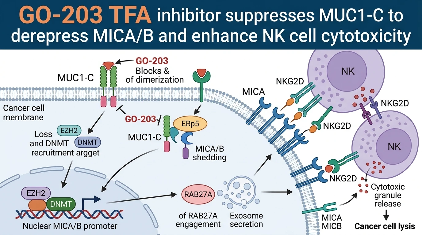
Mucin-1 C-terminal subunit (MUC1-C) functions as a transmembrane signaling protein that coordinates oncogenic drivers and immune evasion programs in epithelial carcinomas. In normal tissues, MUC1 serves protective mucosal functions. Upon oncogenic transformation, MUC1 undergoes aberrant overexpression, cell surface uncoupling, and persistent activation of its C-terminal domain. MUC1-C auto-oligomerizes through Cys-15 residues in its extracellular region. This homo-dimerization is mandatory for its downstream signal transduction. Dimers interact directly with receptor tyrosine kinases, cell survival factors, and transcriptional complexes.
A primary mechanism by which MUC1-C promotes immune evasion involves the systematic down-regulation of natural killer cell receptor group 2, member D (NKG2D) ligands. These ligands, principally MHC class I polypeptide-related sequence A (MICA) and sequence B (MICB), act as molecular beacons for natural killer (NK) cell surveillance. Under physiological stress or malignant transformation, surface MICA/B expression flags abnormal cells for NK cell-mediated lysis. Cancer cells evade this immune surveillance node by suppressing surface MICA/B levels through transcriptional, epigenetic, and post-translational mechanisms.
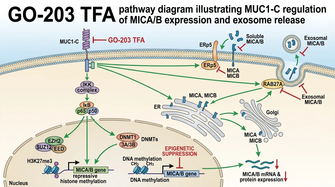
MUC1-C orchestrates a multi-tiered repression mechanism controlling MICA and MICB gene loci. Mechanistically, MUC1-C associates with nuclear factor-kappa B (NF-κB) p65 subunit to drive transcriptional repression cascades. This interaction recruits enhancer of zeste homolog 2 (EZH2), the catalytic subunit of polycomb repressive complex 2 (PRC2). EZH2 catalyzes histone H3 lysine 27 trimethylation (H3K27me3) across MICA/B promoter regions. Simultaneously, MUC1-C recruits DNA methyltransferases (DNMTs), increasing promoter hypermethylation. This combined histone modification and DNA methylation silences MICA/B transcript synthesis, depleting surface ligand availability.
Disruption of this transcriptional suppression is achieved via the synthetic cell-penetrating peptide GO-203. The GO-203 TFA inhibitor targets the Cys-15 motif within the MUC1-C extracellular domain. By directly competing with homodimerization contact points, GO-203 TFA prevents formation of functional MUC1-C signaling complexes. In cell culture models, GO-203 TFA disrupts nuclear MUC1-C assemblies. Consequently, EZH2 and DNMT recruitment to the MICA/B promoters is abrogated. Reversal of chromatin compaction leads to restored MICA and MICB mRNA transcription and surface translation.
Beyond transcriptional silencing, cancer cells frequently down-regulate surface MICA/B through proteolytic shedding and exosomal sequestration. Protein disulfide isomerase ERp5 (PDIA6) facilitates MICA/B cleavage at the cell membrane, shedding soluble MICA/B (sMICA/B) into the tumor microenvironment. sMICA/B acts as a systemic decoy, binding NKG2D receptors on NK cells and triggering receptor internalization. Uniquely, MUC1-C binding to ERp5 promotes this proteolytic cleavage event. GO-203 TFA intervention disrupts the MUC1-C/ERp5 interaction interface, suppressing shedding despite marked ligand upregulation.
Concurrently, MUC1-C engages the small GTPase RAB27A, a key regulator of endosomal trafficking and exosome secretion. MUC1-C forms a direct complex with RAB27A, directing MICA/B into multivesicular bodies for exosomal export. Released MICA/B-bearing exosomes downregulate NKG2D signaling remotely. Treatment with GO-203 TFA disrupts the MUC1-C/RAB27A binding axis. This blockade inhibits exosome release from cancer cells. Retaining MICA and MICB strictly on the plasma membrane converts the tumor cell surface into a potent target for immune clearance. In vitro cytotoxicity assays confirm that GO-203 TFA-treated cells trigger robust NK cell degranulation and tumor killing.
Pharmacological Targeting of MUC1-C and Immune Evasion
Copy code to add this image to your page
GO-203 inhibits MUC1-C dimerization, reversing EZH2- and DNMT-mediated epigenetic silencing of MICA/B ligands. Furthermore, GO-203 disrupts MUC1-C interactions with ERp5 and RAB27A, blocking proteolytic shedding and exosome secretion of MICA/B, respectively. Surface retention of MICA/B markedly enhances NK cell recognition and cytotoxicity against cancer cells.
Derepression of MICA/B Surface Expression via Epigenetic Blockade
MUC1-C signaling maintains repressive chromatin marks across MICA/B loci in human carcinoma cell lines. Nuclear MUC1-C binds directly to NF-κB p65 to assemble a transcriptional silencing complex. This complex recruits EZH2 and DNMT1 to MICA and MICB promoter domains. Chromatin immunoprecipitation assays demonstrate dense H3K27me3 enrichment and focal DNA hypermethylation under baseline conditions. Treatment with GO-203 TFA inhibitor rapidly disrupts MUC1-C nuclear translocation. Within 24 hours of exposure in vitro, promoter-associated EZH2 levels dropped significantly. Decreased H3K27me3 occupancy coincided with elevated MICA/B mRNA expression. Surface MICA and MICB protein levels rose up to four-fold across tested cell lines. The selectivity margin remained intact in non-transformed epithelial cells.
Inhibition of ERp5-Mediated Shedding and RAB27A Exosomal Transport
Proteolytic shedding often counteracts transcriptional induction of MICA/B ligands. MUC1-C physically associates with the thiol oxidoreductase ERp5 at the plasma membrane. This interaction facilitates conformational changes required for ADAM10/17-mediated MICA/B cleavage. Exposure to GO-203 TFA inhibitor blocks MUC1-C/ERp5 complex formation. Soluble sMICA/B concentrations in culture supernatants fell below detection limits following treatment. In parallel, MUC1-C binds directly to RAB27A to drive exosomal sorting of MICA/B. GO-203 TFA treatment abolished RAB27A binding. Vesicular release decreased sharply. Consequently, MICA/B molecules accumulated on the plasma membrane rather than escaping via exosomal pathways.
Potentiation of Natural Killer Cell Mediated Cytotoxic Lysis
Surface accumulation of MICA/B directly restores innate immune recognition. Primary human NK cells co-cultured with peptide-treated tumor cells exhibited heightened activation markers. CD107a degranulation increased significantly in a dose-dependent manner. Interferon-gamma secretion doubled compared to vehicle control arms. Pre-incubation with blocking anti-NKG2D antibodies abrogated this cytotoxicity boost, confirming ligand specificity. Within patient-derived xenograft models, MUC1-C inhibition suppressed tumor growth while augmenting intratumoral NK cell infiltration. These pharmacodynamic observations highlight MUC1-C as a dual therapeutic target for reversing epigenetic silencing and physical evasion mechanisms.