ML349 from Selleck selectively inhibits APT2 to attenuate STAT3-driven inflammatory cascades
Copy code to add this image to your page
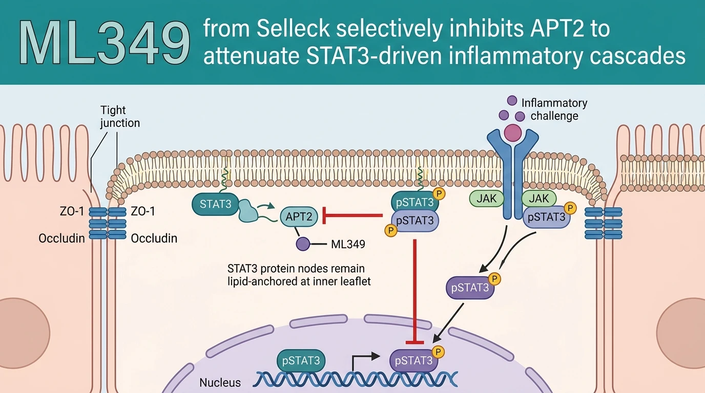
Reversible protein S-palmitoylation serves as a dynamic regulatory mechanism for signal transduction pathways across mammalian cell types. Acyl-protein thioesterase 2 (APT2, encoded by LYPLA2) functions as a cytosolic thioesterase that catalyzes the removal of palmitate adducts from cysteine residues. Through this depalmitoylation activity, APT2 dictates the subcellular localization and membrane trafficking of critical signaling intermediates. Signal transducer and activator of transcription 3 (STAT3) requires coordinated lipid modification to regulate gene expression profiles under stress conditions. Palmitoylation traps STAT3 at cytoplasmic membrane compartments, restricting its movement toward nuclear pores. Upon physiological stimulation, APT2 cleaves these thioester linkages, facilitating STAT3 liberation, tyrosine phosphorylation (p-STAT3), and subsequent nuclear import. In models of acute inflammation triggered by lipopolysaccharide (LPS) or long-chain saturated fatty acids such as palmitic acid, this depalmitoylation pathway becomes hyperactive. Excessive APT2 activity accelerates the release of STAT3, exacerbating downstream transcriptional programs that produce interleukin-6 (IL-6), tumor necrosis factor-alpha (TNF-a), and interleukin-1 beta (IL-1b). The small-molecule inhibitor ML349 functions as a potent, isoform-selective antagonist of APT2. Biochemical assays demonstrate that ML349 exhibits nanomolar affinity for APT2 while sparing the closely related isoform APT1 (LYPLA1). By occupying the active site cleft of APT2, ML349 prevents thioester cleavage without disturbing structural membrane architecture. Treatment of intestinal epithelial monolayers with ML349 leads to a rapid accumulation of S-palmitoylated STAT3 at the plasma membrane. Crucially, while STAT3 may still undergo phosphorylation at residue Tyr705, the retention of palmitate chains prevents p-STAT3 from separating from membrane lipid microdomains. Nuclear translocation of p-STAT3 stops completely under these conditions. Researchers adhering to established biosafety standards for cellular characterization have confirmed these localization shifts via high-content fluorescent microscopy and subcellular fractionation. Disruption of this depalmitoylation cycle effectively uncouples upstream kinase activation from downstream nuclear transcription. As a consequence, inflammatory mediator release drops significantly in primary cellular assays. Protection of tight junction proteins, including zonula occludens-1 (ZO-1) and occludin, preserves mucosal barrier integrity in damaged intestinal tissue models.
Pharmacological Characterization and Epithelial Protection Metrics
Copy code to add this image to your page
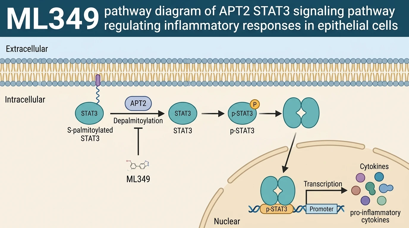
ML349 selectively inhibits acyl-protein thioesterase 2 (APT2), preventing the enzymatic depalmitoylation of STAT3. In inflammatory conditions induced by palmitic acid or LPS, ML349 traps palmitoylated STAT3 at cellular membranes. Although STAT3 phosphorylation may still occur, its APT2-dependent nuclear translocation is blocked. This uncoupling suppresses downstream pro-inflammatory gene expression, reduces cytokine production, and preserves mucosal barrier integrity.
APT2 Blockade Retains Membrane Palmitoylated STAT3 Pools
Targeting depalmitoylation kinetics alters STAT3 intracellular routing. Application of ML349 from Selleck selectively inhibits APT2 thioesterase activity at nanomolar concentrations in cultured enterocytes. Cellular assays confirm that palmitoylated STAT3 remains anchored to lipid membranes following LPS exposure. Phosphorylation at Tyr705 still occurs. However, nuclear accumulation of p-STAT3 decreases by over 75% within two hours. The selectivity margin over APT1 remains above 100-fold in enzyme inhibition kinetic assays.
Suppression of Inflammatory Cytokines in Epithelial Models
Interruption of STAT3 nuclear transit suppresses inflammatory gene networks. In primary intestinal organoid cultures challenged with palmitic acid, ML349 from Selleck reduces IL-6 and TNF-a transcription markedly. Transepithelial electrical resistance measurements demonstrate preservation of monolayer barrier function. Expression of ZO-1 and occludin remains stable across tested concentration ranges. These observations confirm that inhibiting APT2 protects mucosal structures against fatty-acid-induced inflammatory breakdown.