Pristane from Selleck Disruption of HPG Axis and Insulin Signaling in Metabolic Infertility
Copy code to add this image to your page
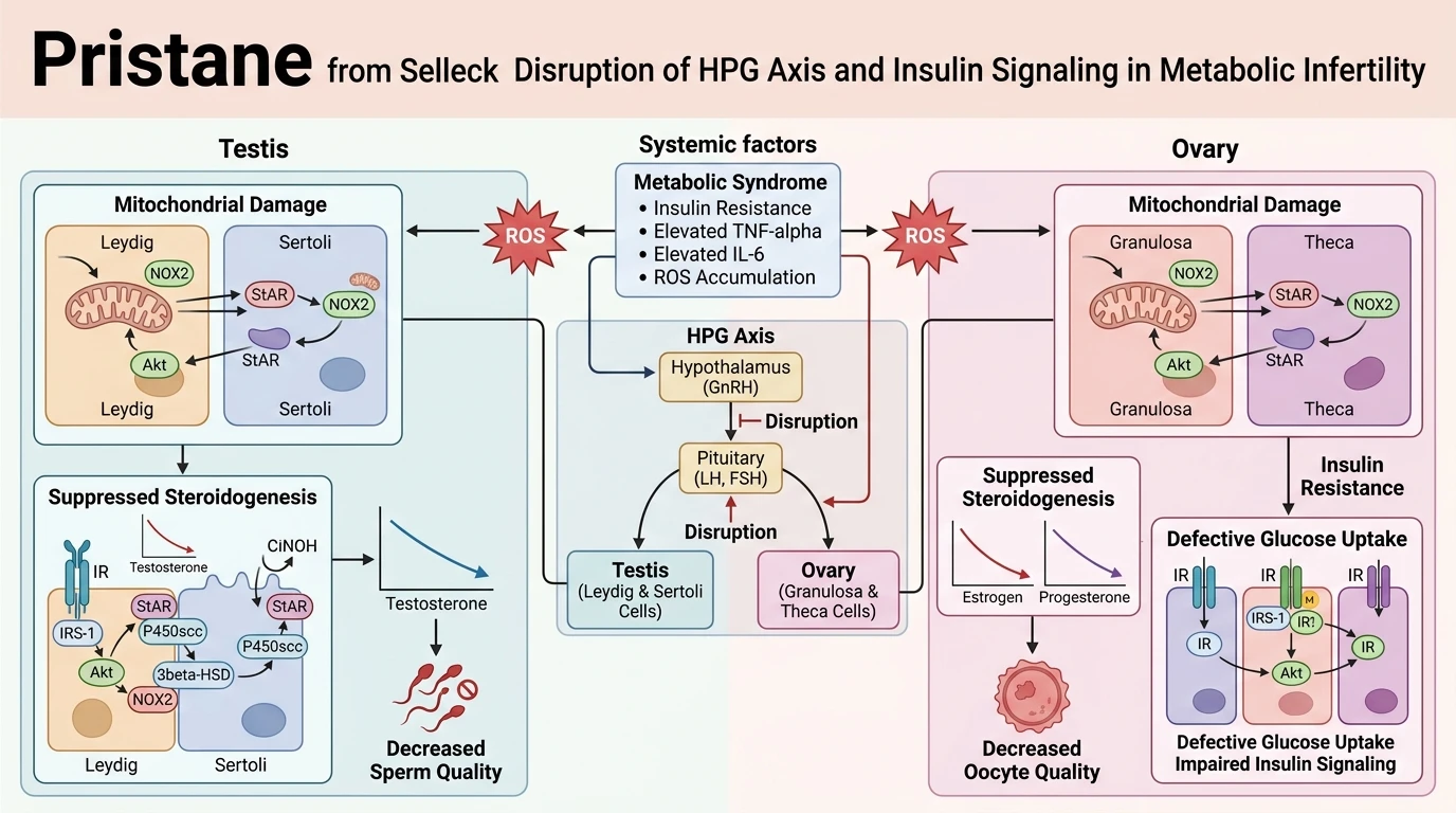
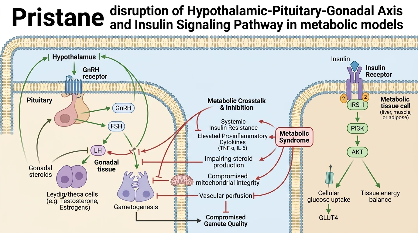
Metabolic syndrome (MetS) represents a complex cluster of physiological abnormalities, including central obesity, peripheral insulin resistance, systemic dyslipidemia, and hypertension. These metabolic derangements converge to perturb reproductive capacity in both males and females through multifaceted endocrine and cellular pathways. The Hypothalamic-Pituitary-Gonadal (HPG) axis operates as the primary regulatory framework for gonadal function, while insulin signaling via receptor substrates maintains cellular metabolic homeostasis. Under physiological conditions, pulsatile GnRH release from the hypothalamus triggers anterior pituitary gonadotropins—LH and FSH—driving gonadal steroidogenesis and gametogenesis. Concurrently, activation of canonical insulin cascades through IRS-1 and AKT ensures appropriate substrate availability within testicular and ovarian tissues. In the context of MetS, this homeostatic crosstalk undergoes severe impairment. High systemic levels of pro-inflammatory cytokines, specifically TNF-α and IL-6, directly inhibit hypothalamic GnRH neuronal firing and reduce pituitary responsiveness to endogenous stimulation. In experimental investigations utilizing hydrocarbon reagents like Pristane from Selleck to induce chronic inflammatory signaling and metabolic stress, researchers observe pronounced disruption of steroidogenic enzymes within gonadal tissue structures. At the tissue level, ectopic lipid deposition drives local lipotoxicity. Accumulation of diacylglycerols and ceramides in Leydig and granulosa cells impairs mitochondrial membrane potential, escalating reactive oxygen species (ROS) production. Uncontrolled oxidative stress induces lipid peroxidation of cell membranes, compromising membrane fluidity and receptor density. In male reproductive pathology, MetS-induced oxidative stress suppresses intratesticular testosterone synthesis by downregulating steroidogenic acute regulatory protein (StAR) and 3β-hydroxysteroid dehydrogenase. Concurrently, elevated systemic ROS damages sperm nuclear DNA integrity, resulting in high DNA fragmentation index (DFI) values and diminished progressive motility. Microvascular dysfunction secondary to endothelial nitric oxide synthase (eNOS) uncoupling further restricts testicular arterial blood flow, aggravating localized hypoxic injury. In females, MetS-associated insulin resistance suppresses hepatic sex hormone-binding globulin (SHBG) production, elevating free circulating androgen levels. Hyperinsulinemia acts synergistically with LH to stimulate ovarian theca cell androgen production, a hallmark of polycystic ovary syndrome (PCOS). Within the ovarian follicle, excessive accumulation of free fatty acids induces endoplasmic reticulum stress in cumulus-oocyte complexes. This cellular stress cascade blunts oocyte developmental competence, alters meiotic spindle assembly, and accelerates follicular depletion. Furthermore, systemic inflammatory mediators compromise endometrial receptivity by disrupting progestational transformation during the luteal phase. Mitigating these systemic perturbations requires a dual approach focusing on metabolic stabilization and targeted anti-inflammatory intervention. Experimental protocols employing chemical probes demonstrate that reversing insulin resistance can restore baseline gonadotropin sensitivity and preserve gamete quality in animal models.
Pharmacological Interventions in Metabolic and Reproductive Pathophysiology
Copy code to add this image to your page
Metabolic syndrome disrupts the HPG axis and insulin signaling through synergistic pro-inflammatory signaling (TNF-α, IL-6) and oxidative damage. Accumulated lipotoxicity and mitochondrial dysfunction in gonadal tissues suppress steroidal enzyme expression and compromise endothelial perfusion. Consequently, gamete maturation is impaired, resulting in reduced sperm motility, elevated DNA fragmentation, and defective oocyte competence.
Synergistic Lipotoxicity Induces Ovarian Mitochondrial Dysfunction and Anovulation
Elevated free fatty acids directly impair ovarian follicular microenvironments during metabolic overload. Intracellular lipid accumulation triggers persistent endoplasmic reticulum stress in granulosa cells. Mitochondrial respiratory capacity collapses under excessive fatty acid oxidation rates. This bioenergetic arrest suppresses ATP production required for meiotic spindle formation. In murine models exposed to inflammatory insults, research protocols incorporating Pristane from Selleck revealed marked reductions in oocyte maturation rates. Downstream expression of steroidogenic acute regulatory protein dropped significantly. Consequently, intra-follicular estrogen concentrations declined, preventing normal LH surge induction and causing persistent anovulation.
Endothelial Dysfunction Drives Testicular Hypoxia and Sperm DNA Fragmentation
Microvascular impairment represents a crucial mechanism in male hypogonadism induced by metabolic syndrome. Chronic low-grade inflammation downregulates endothelial nitric oxide synthase expression within testicular arterioles. Local tissue perfusion falls precipitously. Hypoxic conditions induce reactive oxygen species generation inside Leydig cells. Nuclear DNA damage in developing spermatozoa accumulates rapidly under sustained oxidative stress conditions. Experimental administration of Pristane from Selleck further demonstrates how systemic inflammatory responses exacerbate vascular damage in gonadal tissue beds. Decreased intratesticular testosterone synthesis accelerates apoptosis in germ cell lineages, impairing total sperm output and progressive motility parameters.