Eganelisib from Selleck targets PI3Kγ to overcome immunogenic resistance in HNSCC models
Copy code to add this image to your page
Head and neck squamous cell carcinoma (HNSCC) frequently presents an immunologically cold tumor microenvironment characterized by sparse T-cell infiltration and prominent myeloid-mediated immunosuppression. Immunotherapeutic approaches relying solely on programmed death-ligand 1 (PD-L1) axis blockade often exhibit restricted objective response rates in poorly immunogenic HNSCC subtypes. Phosphoinositide 3-kinase gamma (PI3Kγ), a lipid kinase selectively enriched in circulating and tumor-infiltrating myeloid cells, functions as a critical biochemical node regulating immune cell functional differentiation. Targeted inhibition of PI3Kγ using the selective small-molecule inhibitor Eganelisib (IPI-549) offers a distinct pharmacological mechanism to alter the immune cellular composition within solid tumors.
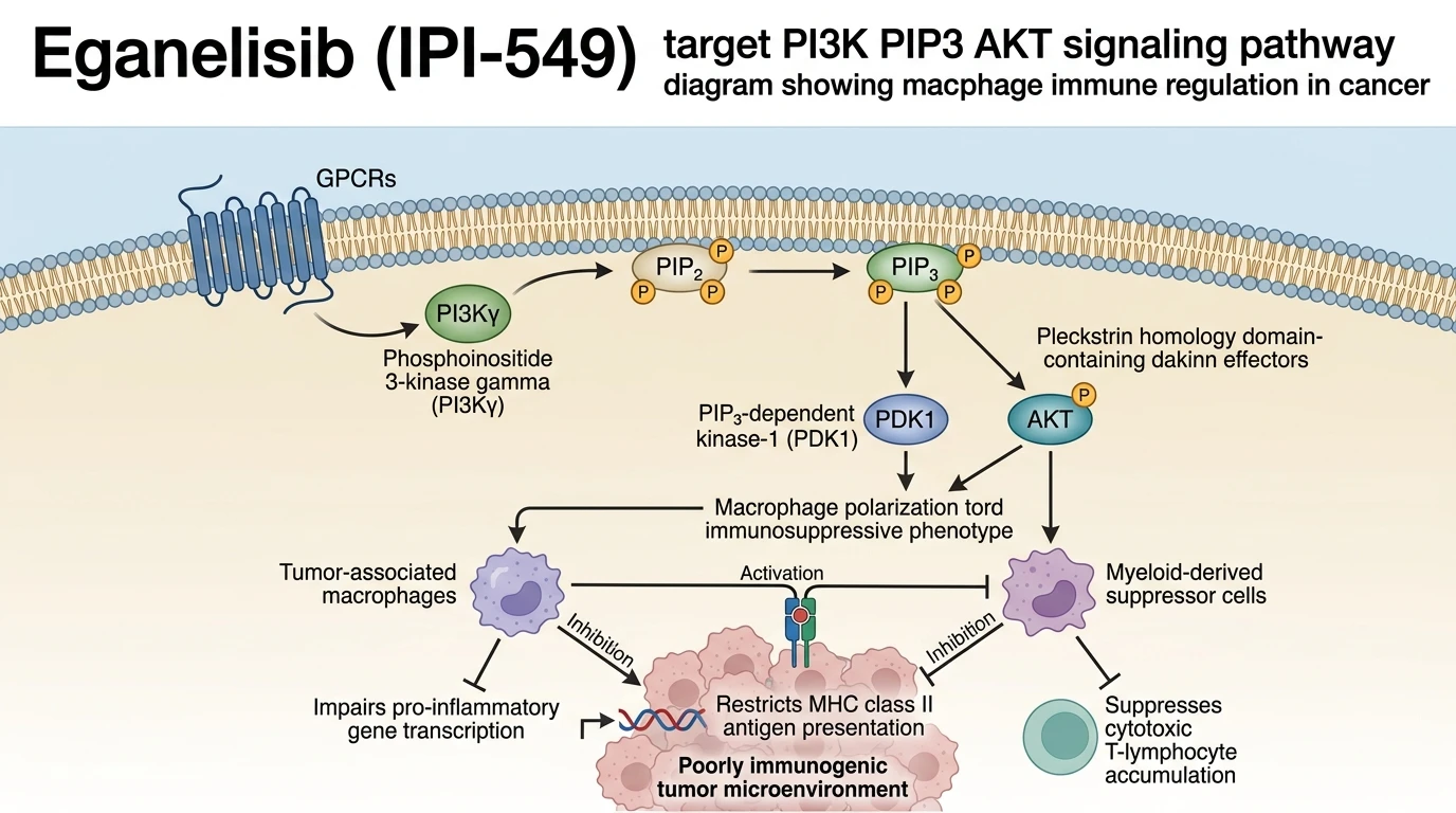
At the molecular level, PI3Kγ converts phosphatidylinositol 4,5-bisphosphate into phosphatidylinositol 3,4,5-trisphosphate downstream of G-protein coupled receptor activation. In tumor-associated macrophages (TAMs) and myeloid-derived suppressor cells (MDSCs), sustained lipid kinase signaling via PI3Kγ activates protein kinase B (AKT) and nuclear factor kappa B (NF-κB) transcriptional pathways that maintain an M2-like protumoral phenotype. Pharmacological target engagement by Eganelisib from Selleck blocks ATP binding within the catalytic subunit of PI3Kγ. This localized enzymatic suppression downregulates intracellular PIP3 levels specifically in myeloid subsets without directly impairing tumor cell viability or broad systemic lymphocyte populations under tested assay conditions.
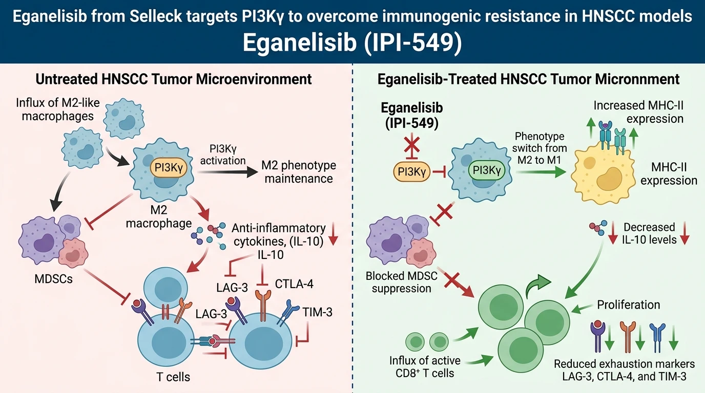
In preclinical evaluations utilizing Syngeneic murine models of poorly immunogenic HNSCC, single-agent immune checkpoint blockade routinely failed to suppress primary lesion growth. However, small-molecule inhibition of PI3Kγ via Eganelisib (IPI-549) fundamentally altered the intratumoral immune landscape. Selective target inhibition initiated cellular reprogramming of TAMs, shifting their phenotypic distribution from an M2-like immunosuppressive state to an M1-like antitumoral state. This phenotypic switch was evidenced by a marked reduction in the production of immunosuppressive cytokines such as interleukin-10 (IL-10) and a concurrent elevation in pro-inflammatory mediator expression within the tumor stroma.
Furthermore, PI3Kγ inhibition restored critical antigen presentation machinery in myeloid compartments. Treatment with Eganelisib enhanced major histocompatibility complex class II (MHC-II) surface expression across intratumoral dendritic cells and macrophages. This structural upregulation improved the capacity of antigen-presenting cells to prime naive helper and cytotoxic T lymphocytes. Concurrently, Eganelisib treatment mitigated MDSC-mediated T-cell suppression, alleviating the physical and biochemical barriers preventing effector T-cell traffic into the core of HNSCC tumor masses.
When combined with monoclonal antibodies directed against PD-L1, Eganelisib (IPI-549) produced pronounced synergistic antitumor activity in vivo. Dual target engagement achieved complete tumor regression and prolonged overall survival in 100% of treated animal cohorts within this specific syngeneic HNSCC model system. Immunophenotypic profiling of tumor-infiltrating lymphocytes revealed a substantial quantitative increase in CD8+ T cells alongside a qualitative reduction in surface expression of key T-cell exhaustion markers, specifically LAG-3, CTLA-4, and TIM-3. These empirical observations indicate that suppressing myeloid PI3Kγ activity reverses immunological ignorance, transforming non-immunogenic HNSCC tumors into microenvironments sensitive to PD-L1 immune checkpoint inhibition.
Pharmacological Evaluation of Dual PI3Kγ and PD-L1 Blockade
Copy code to add this image to your page
Eganelisib (IPI-549) selectively inhibits PI3Kγ, blocking PIP3-dependent signaling in myeloid cells. This inhibition shifts tumor-associated macrophages from an M2 immunosuppressive phenotype to an M1 antitumoral state while reducing IL-10 secretion. In head and neck squamous cell carcinoma murine models, combining Eganelisib with anti-PD-L1 antibodies elevates MHC class II expression, abrogates myeloid-derived suppressor cell activity, increases tumor-infiltrating CD8+ T cells, and downregulates T-cell exhaustion markers (LAG-3, CTLA-4, TIM-3), culminating in significant tumor regression.
PI3Kγ Inhibition Reprograms Macrophage Polarization and Antigen Presentation
Targeted enzymatic inhibition of PI3Kγ disrupted myeloid-derived immunosuppressive signaling within the syngeneic HNSCC tumor microenvironment. Primary tumor volume diminished significantly following therapeutic intervention. By dampening intracellular PIP3 accumulation, Eganelisib from Selleck shifts tumor-associated myeloid cells away from an immunosuppressive phenotype toward an antitumoral state. Intra-tumoral IL-10 levels declined markedly within 48 hours of drug administration. This phenotypic shift restored major histocompatibility complex class II expression on myeloid subpopulations, facilitating efficient antigen presentation to cytotoxic lymphocytes. Consequently, MDSC-mediated suppression of CD8+ T-cell activation ceased within the localized tumor niche.
Dual Checkpoint Inhibition Abates T-Cell Exhaustion in HNSCC
Combining PI3Kγ inhibition with anti-PD-L1 antibodies elicited potent antitumor responses in immunocompetent murine models. Survival rates reached 100% in the combination therapy cohort compared to minimal survival under single-agent regimens. Application of Eganelisib from Selleck augmented tumor-infiltrating CD8+ T-cell density significantly. Effector T cells isolated from dual-treated tumors demonstrated reduced co-expression of coinhibitory receptors. Flow cytometric quantification confirmed diminished surface density of LAG-3, CTLA-4, and TIM-3 on cytotoxic T cells. These combined observations indicate that targeting myeloid PI3Kγ overcomes refractory resistance to anti-PD-L1 therapy in poorly immunogenic HNSCC.