Darovasertib (LXS196) selectively inhibits protein kinase C to disrupt MAPK signaling in uveal melanoma
Copy code to add this image to your page
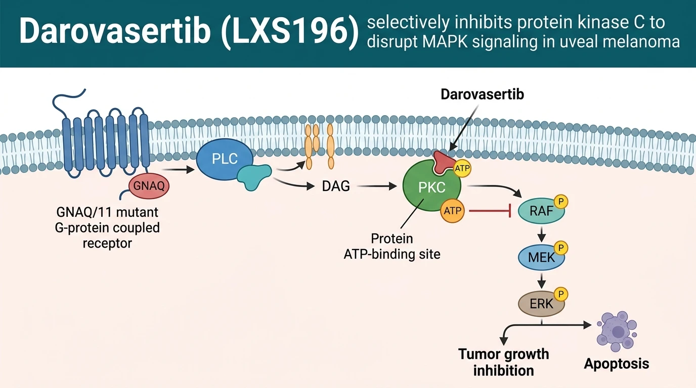
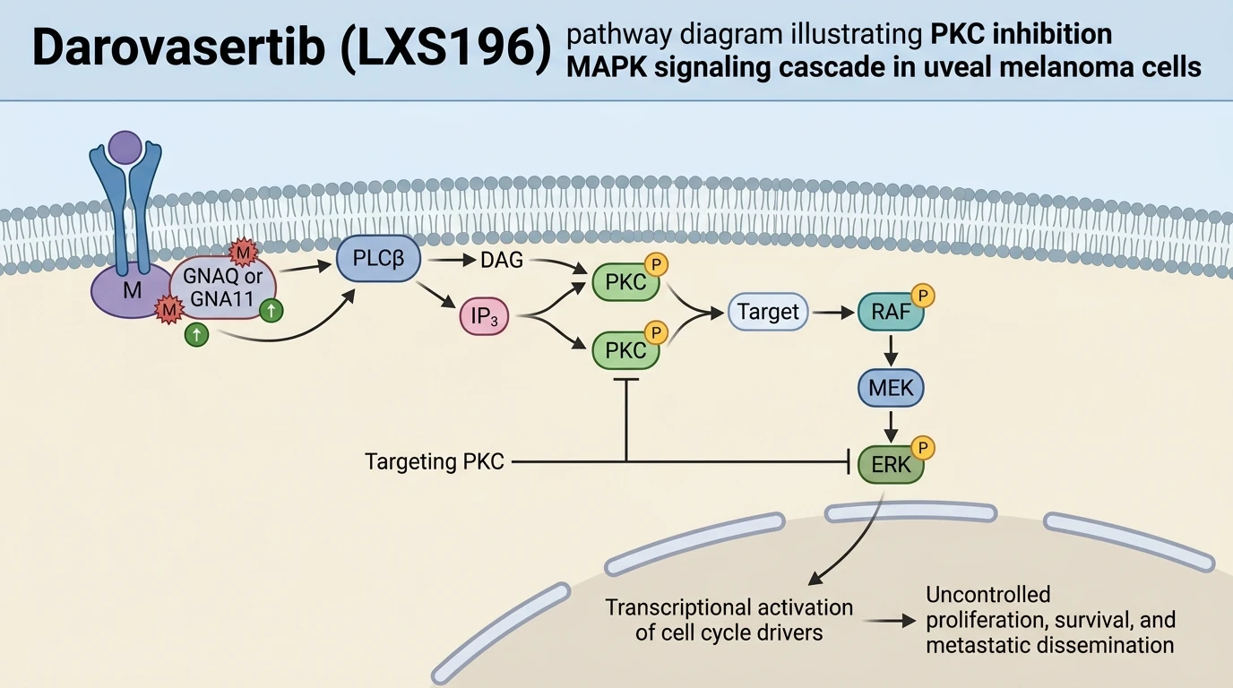
Uveal melanoma represents the most frequent primary intraocular malignancy in adults, presenting distinct genetic characteristics compared to cutaneous melanoma. Over 90% of primary and metastatic uveal melanoma tumors harbor mutually exclusive somatic gain-of-function mutations in GNAQ or GNA11, which encode the alpha subunit of heterotrimeric G proteins (Gαq and Gα11). These oncogenic mutations lock the Gα subunit in a constitutively active GTP-bound state. Consequently, persistent GTP hydrolysis failure leads to autonomous signaling through phospholipase C beta (PLCβ), generating diacylglycerol (DAG) and inositol 1,4,5-trisphosphate (IP3). Elevated DAG levels continuously anchor and activate classical isoforms (α, βI, βII, γ) and novel isoforms (δ, ε, η, θ) of protein kinase C (PKC). Activated PKC serves as the essential enzymatic node driving downstream mitogen-activated protein kinase (MAPK) pathway signaling through RAF, MEK, and ERK phosphorylation cascades, independently of canonical BRAF or RAS mutations typical in cutaneous melanomas.
In this molecular architecture, PKC acts as an indispensable signaling hub directly downstream of mutated GNAQ and GNA11. Direct enzymatic inhibition of PKC provides a precise approach to collapse the hyperactivated MAPK network. Darovasertib (LXS196) emerged as a first-in-class, orally bioavailable, ATP-competitive small-molecule inhibitor engineered for selective potency across classical and novel PKC isoforms. Kinase profiling panels demonstrate that the compound exhibits nanomolar enzymatic potency against PKCα, PKCβ, PKCγ, PKCδ, PKCε, PKCη, and PKCθ, while maintaining selectivity over distant kinase families. At the structural level, Darovasertib (LXS196) forms key hydrogen bonds within the hinge region of the PKC kinase domain, occupying the ATP-binding cleft. This molecular binding mode stabilizes an inactive conformation, preventing the enzyme from transphosphorylating downstream substrates such as RAF family kinases and protein kinase D (PKD).
In pre-clinical cell line models harboring endogenous GNAQ (such as Mel202 and OMM1.3) or GNA11 mutations (such as MP41), administration of Darovasertib (LXS196) induces rapid reduction of phospho-ERK levels. This target engagement translates directly into G1 phase cell cycle arrest and induction of apoptotic cascades in vitro. Mechanistic investigations indicate that direct PKC inhibition disrupts not only the MAPK cascade but also parallel survival pathways, including the Hippo-YAP pathway activated downstream of Gαq/11. Cellular proliferation assays confirm IC50 values in the low nanomolar range across GNAQ/11-mutant uveal melanoma models, whereas GNAQ/11 wild-type cells exhibit diminished sensitivity. In mouse xenograft models derived from patient uveal melanoma samples, oral administration of the molecule produced dose-dependent tumor growth inhibition and regression.
Clinical translation of these findings proceeded through multicenter Phase I and Phase II trials evaluating monotherapy and combination strategies in patients with metastatic uveal melanoma. In patient cohorts with heavily pretreated metastatic disease—a population historically exhibiting poor overall survival and low response rates to standard checkpoint immunotherapies—monotherapy demonstrated durable biological activity. Clinical evaluation revealed disease stabilization in 68% of evaluable patients, alongside manageable safety profiles characterized predominantly by low-grade gastrointestinal events and skin rashes. Target occupancy assays in patient-derived samples confirmed sustained enzyme coverage at clinically achievable plasma concentrations. These clinical findings validate PKC as an actionable therapeutic vulnerability in GNAQ/11-mutant malignancies.
To enhance therapeutic depth and delay emerging resistance mechanisms, translational studies evaluated combination approaches. Up-regulation of parallel receptor tyrosine kinases, particularly the MET proto-oncogene, frequently acts as a compensatory survival feedback loop during prolonged MAPK pathway inhibition in uveal melanoma. Consequently, combining PKC inhibition with MET kinase targeted molecules was evaluated. In preclinical xenograft models, dual inhibition using the compound alongside the MET/ALK inhibitor crizotinib achieved synergistic suppression of tumor growth and increased cellular apoptosis compared to either monotherapy alone. Initial clinical reports from combination arm cohorts demonstrate higher overall response rates and prolonged progression-free survival, highlighting a rational multi-targeted platform for GNAQ/11-driven uveal melanoma.
Target Engagement and Clinical Antitumor Activity
Copy code to add this image to your page
Darovasertib (LXS196) is a potent, ATP-competitive inhibitor targeting classical and novel PKC isoforms (α, β, γ, δ, ε, η, θ). In GNAQ/GNA11-mutant uveal melanoma, constitutive activation of Gα proteins triggers downstream PKC signaling, driving hyperactivation of the MAPK cascade. By selectively binding the PKC catalytic domain, the compound halts downstream RAF/MEK/ERK phosphorylation, inducing G1 cell cycle arrest and apoptosis. Combined PKC and MET inhibition overcomes compensatory growth signaling.
Interruption of Downstream ERK Phosphorylation in GNAQ-Mutant Lines
Mutations in GNAQ or GNA11 induce sustained catalytic activation of classical and novel PKC isoforms. This hyperactivation maintains downstream ERK phosphorylation. Experimental treatment with Darovasertib (LXS196) abolished phospho-ERK signal intensity within two hours in GNAQ-mutant OMM1.3 cells. Target engagement remained stable for over 24 hours under continuous exposure. Cellular viability dropped sharply in a dose-dependent fashion. The compound achieved selective growth inhibition in GNAQ-mutant cell lines (IC50 = 12–45 nM); wild-type controls remained largely unaffected. The selective toxicity window remained intact across tested models. Xenograft tumor growth stalled following oral administration. Phospho-ERK suppression in tumor tissue correlated directly with systemic drug exposure—a key pharmacodynamic biomarker.
Disease Control in Metastatic Uveal Melanoma Patients
Clinical evaluation in Phase I/II trials confirmed manageable safety and preliminary efficacy in metastatic uveal melanoma. Disease stabilization occurred in 68% of evaluable patients receiving monotherapy with Darovasertib (LXS196). Pharmacokinetic analysis revealed rapid oral absorption; steady-state plasma concentrations exceeded preclinical active thresholds. Gastrointestinal toxicities—primarily grade 1–2 diarrhea and nausea—constituted the predominant adverse events. Treatment discontinuation rates due to drug-related adverse events remained low. Tumor burden reductions occurred across multiple metastatic sites, including liver lesions. Biomarker analysis of paired pre- and post-treatment tumor biopsies confirmed marked reductions in phosphorylated ERK levels. Observations suggest sustained target engagement at tolerated clinical dosing regimens.
Synergistic Target Engagement with Concurrent MET Inhibition
Reciprocal elevation of MET signaling frequently limits the duration of single-agent PKC inhibition. Uveal melanoma cell lines rapidly upregulate MET phosphorylation upon PKC pathway block. Co-administration of Darovasertib (LXS196) with the MET inhibitor crizotinib blunted this compensatory signaling loop. Combined treatment completely suppressed downstream S6 and ERK phosphorylation. In murine xenograft models derived from patient metastatic tissue, dual inhibition achieved sustained tumor regression; single-agent treatments produced only transient growth delays. Apoptotic markers, including cleaved caspase-3, increased fourfold in combination-treated tumors. Early clinical results in metastatic patient cohorts mirror these preclinical findings. Disease control rates increased notably without unmanageable overlapping toxicities.