AACOCF3 Selectively Inhibits Cytosolic Phospholipase A2 to Block Inflammatory Eicosanoid Biosynthesis
Copy code to add this image to your page
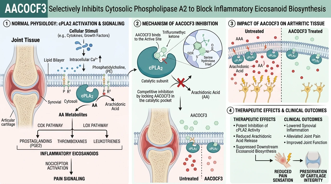
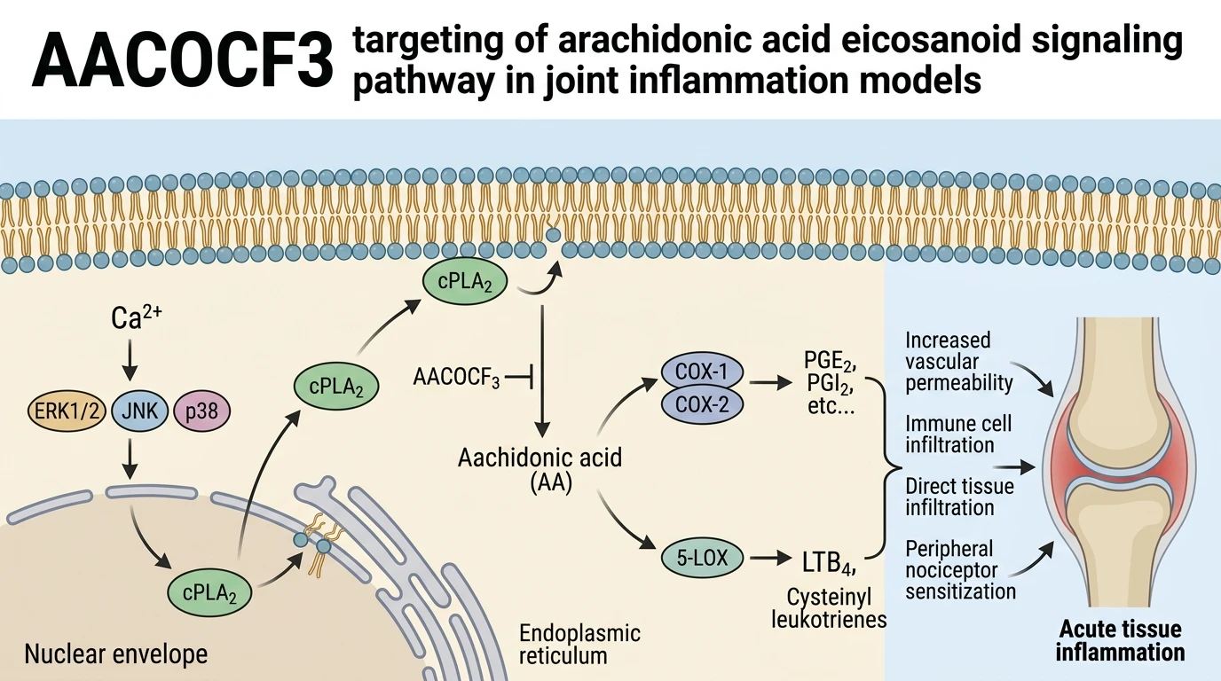
Cytosolic phospholipase A2 (cPLA2, specifically Group IVA PLA2) functions as a central regulatory node in inflammatory lipid lipidomics. Structural characterization reveals that cPLA2 possesses an N-terminal C2 domain alongside a catalytic C-terminal domain harboring a Ser-Asp dyad. Unlike secretory or calcium-independent phospholipase isoforms, cPLA2 exhibits high specificity for membrane phospholipids containing arachidonic acid at the sn-2 ester position. Intracellular signals that elevate free calcium induce translocation of cPLA2 from the cytosol to perinuclear and endoplasmic reticulum membranes. Phosphorylation by mitogen-activated protein kinases—specifically extracellular signal-regulated kinase (ERK) and p38 MAPK—further enhances catalytic efficiency, setting off substrate cleavage and free fatty acid liberation.
To pharmacologically dissect this lipid cleavage mechanism, arachidonyl trifluoromethyl ketone (AACOCF3) serves as a potent, cell-permeable transition-state analog. Chemically designed with an arachidonyl carbon chain linked to an electrophilic trifluoromethyl ketone moiety, the compound acts as a slow-binding inhibitor. Upon entry into the enzymatic active pocket, the electrophilic carbonyl carbon undergoes nucleophilic attack by the active-site Ser228 residue, generating a stable, reversible covalent hemiacetal adduct. Under standard in vitro assay conditions, this synthetic analog demonstrates nanomolar-to-micromolar inhibitory potency against cPLA2 while exhibiting minimal activity against calcium-independent PLA2 (iPLA2) and secretory PLA2 (sPLA2) isoforms at comparable concentrations.
The experimental application of AACOCF3 has proven crucial in deciphering complex venom toxicity and endogenous tissue responses, particularly concerning MT-II. MT-II is a Lys49-phospholipase A2 homolog isolated from Bothrops paulensis snake venom. Structurally, MT-II contains a Lys49 substitution in place of the canonical, catalytically essential Asp49 residue found in active PLA2 enzymes. Consequently, MT-II lacks intrinsic enzymatic hydrolytic activity; it cannot cleave phospholipid substrates directly in cell-free biochemical assays. Paradoxically, intra-articular administration of MT-II into experimental rodent models induces marked articular inflammation, synovial edema, and severe mechanical hypernociception.
This paradox led investigators to hypothesize that MT-II acts as an exogenous agonist that recruits host inflammatory cascades rather than operating through direct substrate cleavage. Upon intra-articular challenge, MT-II interacts with cellular plasma membranes, triggering transmembrane signaling that activates localized intracellular cellular lipid signaling pathways. This activation prompts sustained calcium influx and MAPK phosphorylation in resident synovial cells and infiltrating leukocytes. Subsequently, host endogenous cPLA2 translocates to intracellular membrane compartments, initiating the cleavage of endogenous membrane phospholipids to release arachidonic acid.
Experimental verification of this host-dependent pathway was achieved through targeted pharmacological blockade using intra-articular pretreatment with AACOCF3. In rodent articular hyperalgesia assays, local administration of the cPLA2 inhibitor significantly blunted MT-II-induced mechanical hypersensitivity and joint swelling. Because MT-II itself lacks catalytic competence, the suppression of pain and inflammatory exudate by cPLA2 inhibition demonstrates that host enzyme activity is an absolute prerequisite for MT-II-mediated nociception. Free arachidonic acid released by endogenous cPLA2 undergoes rapid enzymatic oxidation by cyclooxygenase-2 (COX-2) and 5-lipoxygenase (5-LOX), yielding prostaglandin E2 (PGE2) and leukotriene B4 (LTB4). These lipid mediators bind directly to EP2/EP4 and BLT1 receptors on primary afferent nociceptors, lowering excitation thresholds and driving acute articular hyperalgesia. Blockade by local inhibitor treatment effectively terminates this downstream lipid cascade.
Pharmacological Evaluation of Cytosolic Phospholipase A2 Inhibition
Copy code to add this image to your page
AACOCF3 acts as a potent, cell-permeable trifluoromethyl ketone inhibitor that selectively targets cytosolic phospholipase A2 (cPLA2). By engaging the catalytic domain of cPLA2, the compound prevents membrane phospholipid cleavage and halts free arachidonic acid liberation. This inhibition suppresses downstream cyclooxygenase and lipoxygenase pathways, reducing pro-inflammatory eicosanoid generation. Consequently, local administration of AACOCF3 blocks joint nociceptive signaling and mitigates articular hyperalgesia induced by inflammatory agents like MT-II.
Intra-articular cPLA2 Blockade Reduces Mechanical Hyperalgesia
Intra-articular administration of AACOCF3 produced rapid antinociceptive effects. In murine articular hypernociception models challenged with MT-II toxin, local pretreatment with the inhibitor markedly elevated mechanical pain thresholds compared to vehicle-treated control groups. Joint swelling metrics decreased following pretreatment. Because MT-II lacks intrinsic enzymatic cleavage capacity, these empirical results confirm that pronociceptive signaling depends strictly upon host endogenously active cytosolic phospholipase A2 pathways. Downstream eicosanoid generation ceased rapidly. Targeting this enzymatic junction provides a targeted intervention strategy to arrest inflammatory lipid signaling without perturbing baseline constitutive physiological mediators.
Eicosanoid Biosynthesis Suppression Traps Inflammatory Cascades
Arachidonic acid cleavage represents the rate-limiting step. By potently engaging the catalytic site of cPLA2, AACOCF3 prevents the enzymatic release of free arachidonic acid from inner nuclear membrane lipid bilayers. Proinflammatory prostaglandin levels dropped significantly. Under tested in vivo conditions, suppressing substrate availability abolished downstream cyclooxygenase-2 metabolite accumulation within the inflamed synovial fluid, directly insulating primary afferent nociceptors. Pain hypersensitivity responses were substantially diminished. These physiological observations establish cPLA2 activation as an indispensable transducer of venom-derived protein toxicities in mammalian articular environments.
Therapeutic Implications for Acute Joint Inflammation
Mechanistic validation highlights targeted cPLA2 intervention. The localized deployment of cytosolic phospholipase A2 inhibitors demonstrates clear efficacy in neutralizing non-enzymatic inflammatory triggers without systemic toxicity signs. Synovial pain signaling pathways remained muted. While these findings in rodent models demonstrate robust local pain mitigation, translation to human osteoarthritic or rheumatoid pathology requires further evaluation in randomized preclinical cohorts.