Bleomycin hydrochloride induces DNA damage and pulmonary fibrosis mediated by TPH1-dependent serotonin signaling
Copy code to add this image to your page
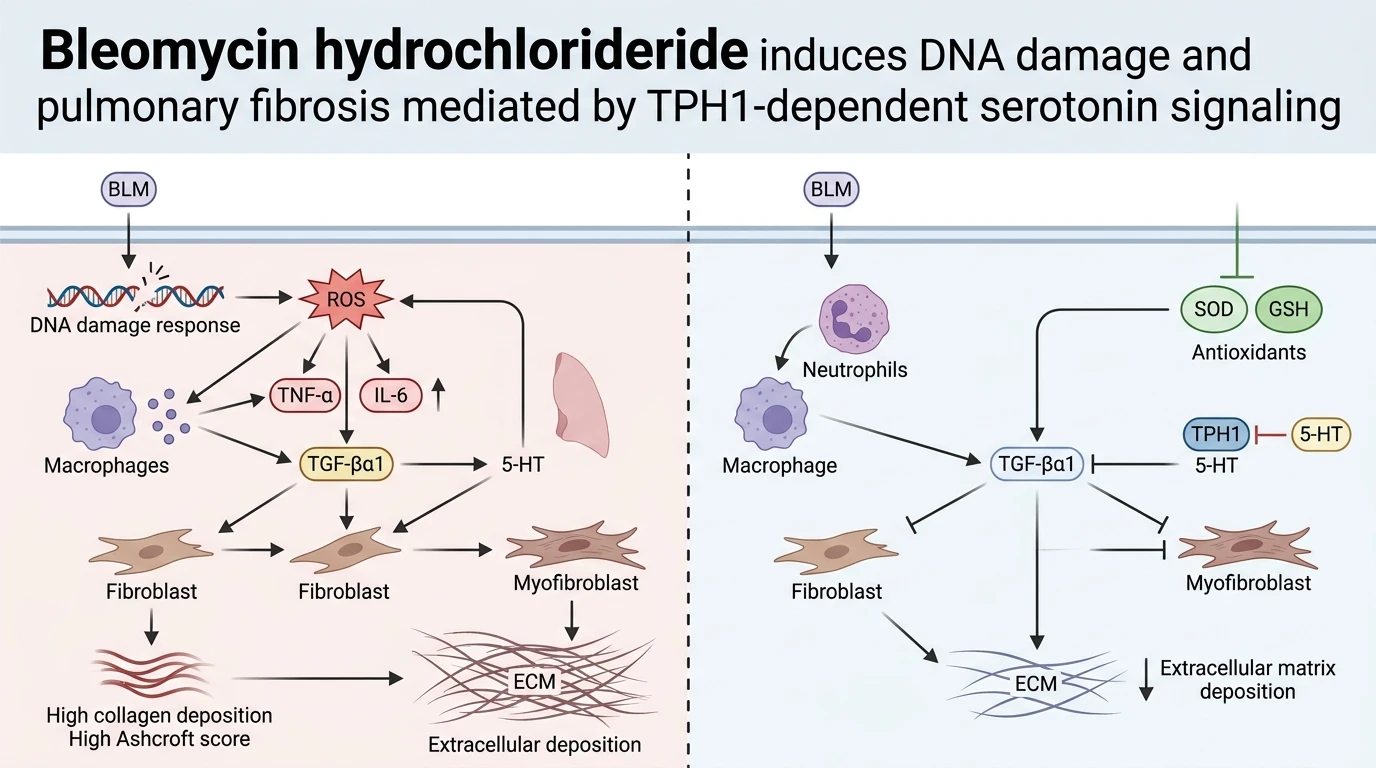
Intratracheal administration of Bleomycin hydrochloride remains a benchmark experimental model for evaluating acute lung injury and subsequent parenchymal pulmonary fibrosis. At the molecular level, Bleomycin hydrochloride forms an iron-dependent active complex that reacts with molecular oxygen. This reaction generates reactive oxygen species, directly causing single- and double-stranded DNA cleavage in pulmonary alveolar epithelial cells. The resulting genomic instability initiates cellular apoptosis and necrosis. Alveolar barrier breakdown follows immediately. Subsequent tissue repair mechanisms frequently become dysregulated, precipitating persistent inflammatory cell influx and uncontrolled collagenous matrix deposition.
Recent investigation examined whether peripheral serotonin (5-HT)—synthesized primarily by the rate-limiting enzyme tryptophan hydroxylase 1 (TPH1)—serves as a crucial biochemical amplifier in this cascade. To determine the functional role of non-neural 5-HT, researchers subjected wild-type (WT) and TPH1 knockout (KO) mice to single-dose intratracheal Bleomycin hydrochloride challenge. The experimental outcome established a striking divergence in disease progression between cohorts. Wild-type mice developed rapid systemic weight loss, profound intra-alveolar exudation, and extensive fibrotic consolidation. Conversely, TPH1 KO animals displayed marked protection across multiple pathophysiological parameters.
Morphological and histological evaluations corroborated the protective phenotype of TPH1 deficiency. Lung tissues from WT mice exposed to Bleomycin hydrochloride exhibited severe alveolar wall thickening, widespread architectural disruption, and extensive fibroblastic foci formation. Ashcroft fibrotic scores were dramatically elevated in WT lungs. In contrast, TPH1 KO mice retained preserved alveolar architecture and significantly lower Ashcroft scores. Quantitative measurement of pulmonary collagen content via hydroxyproline assay demonstrated reduced collagen accumulation in KO lung tissues relative to WT controls. These histological observations indicate that peripheral serotonin signaling directly supports the fibrotic transition within the pulmonary parenchyma.
Analysis of bronchoalveolar lavage fluid (BALF) provided critical insights into vascular permeability and leukocyte recruitment. Intratracheal Bleomycin hydrochloride administration in WT mice triggered substantial BALF total protein exudation and massive inflammatory cell infiltration. Neutrophil recruitment was especially prominent. Myeloperoxidase (MPO) activity assays confirmed abundant neutrophil accumulation within WT lung tissues. However, TPH1 KO mice exhibited markedly attenuated BALF protein exudation and diminished total cell counts. MPO-positive cell counts remained significantly depressed in KO lungs, demonstrating that peripheral 5-HT signaling promotes inflammatory cell extravasation into damaged alveolar spaces.
The inflammatory profile was further characterized by measuring key pro-inflammatory cytokines. In WT lung homogenates, Bleomycin hydrochloride insult triggered robust upregulation of tumor necrosis factor-alpha (TNF-alpha) and interleukin-6 (IL-6). TPH1 KO mice exhibited significantly blunted TNF-alpha and IL-6 expression levels. Because TNF-alpha and IL-6 recruit secondary immune effectors and sustain myofibroblast activation, their attenuation in TPH1 KO mice underlines a major mechanism of reduced pathology.
Oxidative stress parameters further revealed the cellular impact of 5-HT deficiency. Lung tissue from WT mice displayed elevated malondialdehyde (MDA) levels alongside reduced superoxide dismutase (SOD) activity and depleted glutathione (GSH) reserves. This imbalance indicates severe lipid peroxidation and antioxidant defense exhaustion following Bleomycin hydrochloride exposure. In TPH1 KO mice, MDA accumulation was markedly diminished. Endogenous SOD activity and GSH concentrations were substantially preserved. These results suggest that 5-HT amplifies tissue damage partly by driving oxidative exhaustion.
At the transcript and protein level, expression of collagen type I and transforming growth factor-beta 1 (TGF-beta1) was strongly suppressed in TPH1 KO lungs compared to WT counterparts. TGF-beta1 operates as the primary pro-fibrotic cytokine driving epithelial-mesenchymal transition and extracellular matrix synthesis. The downregulation of TGF-beta1 in TPH1 KO mice directly correlates with decreased collagen I transcription and attenuated tissue remodeling. Empirical observations demonstrate that peripheral 5-HT signaling exacerbates Bleomycin hydrochloride-induced pulmonary fibrosis through coordinated enhancement of oxidative stress, leukocyte infiltration, inflammatory cytokine release, and TGF-beta1-driven extracellular matrix synthesis.
Pathophysiological Mechanisms and Serotoninergic Modulation of Lung Fibrosis
Copy code to add this image to your page
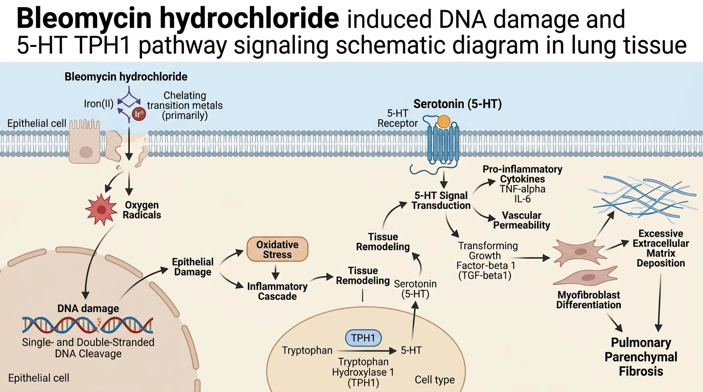
Bleomycin hydrochloride induces single- and double-stranded DNA breaks, generating reactive oxygen species and triggering alveolar epithelial injury. This insult activates an inflammatory cascade characterized by neutrophil infiltration, lipid peroxidation, and cytokine secretion. Peripheral serotonin, synthesized via TPH1, amplifies this pathology by promoting TNF-alpha and IL-6 release, accelerating antioxidant depletion, and upregulating TGF-beta1. TGF-beta1 subsequently drives collagen I synthesis and myofibroblast activation, leading to extracellular matrix deposition and pulmonary fibrosis. TPH1 deficiency attenuates these deleterious processes, demonstrating that 5-HT signaling exacerbates bleomycin-induced fibrogenesis.
Attenuated Inflammatory Cell Recruitment in TPH1-Deficient Lung Tissue
Intratracheal instillation of Bleomycin hydrochloride triggers acute epithelial disruption and leukocyte accumulation. Neutrophils rapidly infiltrate alveolar spaces following initial insult. In wild-type mice, this inflammatory response remains sustained over several days. Infiltrated cells release myeloperoxidase and pro-inflammatory cytokines. In contrast, TPH1 knockout mice show reduced cell influx. BALF total protein levels remained low in TPH1 KO animals under identical challenge conditions. Neutrophil density was notably reduced. Furthermore, pulmonary TNF-alpha and IL-6 protein concentrations decreased significantly. These empirical metrics confirm that peripheral serotonin acts as an upstream driver of pulmonary leukocyte migration within this murine model.
Preserved Antioxidant Defense and Downregulated Fibrotic Signal Transduction
Oxidative damage represents a primary pathway of pulmonary tissue degradation. Following exposure to Bleomycin hydrochloride, wild-type lung tissue accumulated high concentrations of malondialdehyde. Endogenous antioxidant reserves suffered rapid depletion. SOD activity dropped. Glutathione levels declined sharply. Conversely, TPH1 knockout mice maintained endogenous SOD activity and glutathione pools. Lipid peroxidation markers were low. Additionally, expression of TGF-beta1 and collagen type I fell substantially in TPH1 KO lungs. Downstream myofibroblast activation ceased to progress. Lower Ashcroft scores and reduced hydroxyproline content confirmed diminished extracellular matrix accumulation. Thus, targeting peripheral TPH1-mediated serotonin synthesis offers a viable strategy to suppress TGF-beta1-driven matrix turnover.