AP-III-a4 (ENOblock) Others inhibitor targeting enolase suppresses RhoA ROCK signaling and neuroinflammation
Copy code to add this image to your page
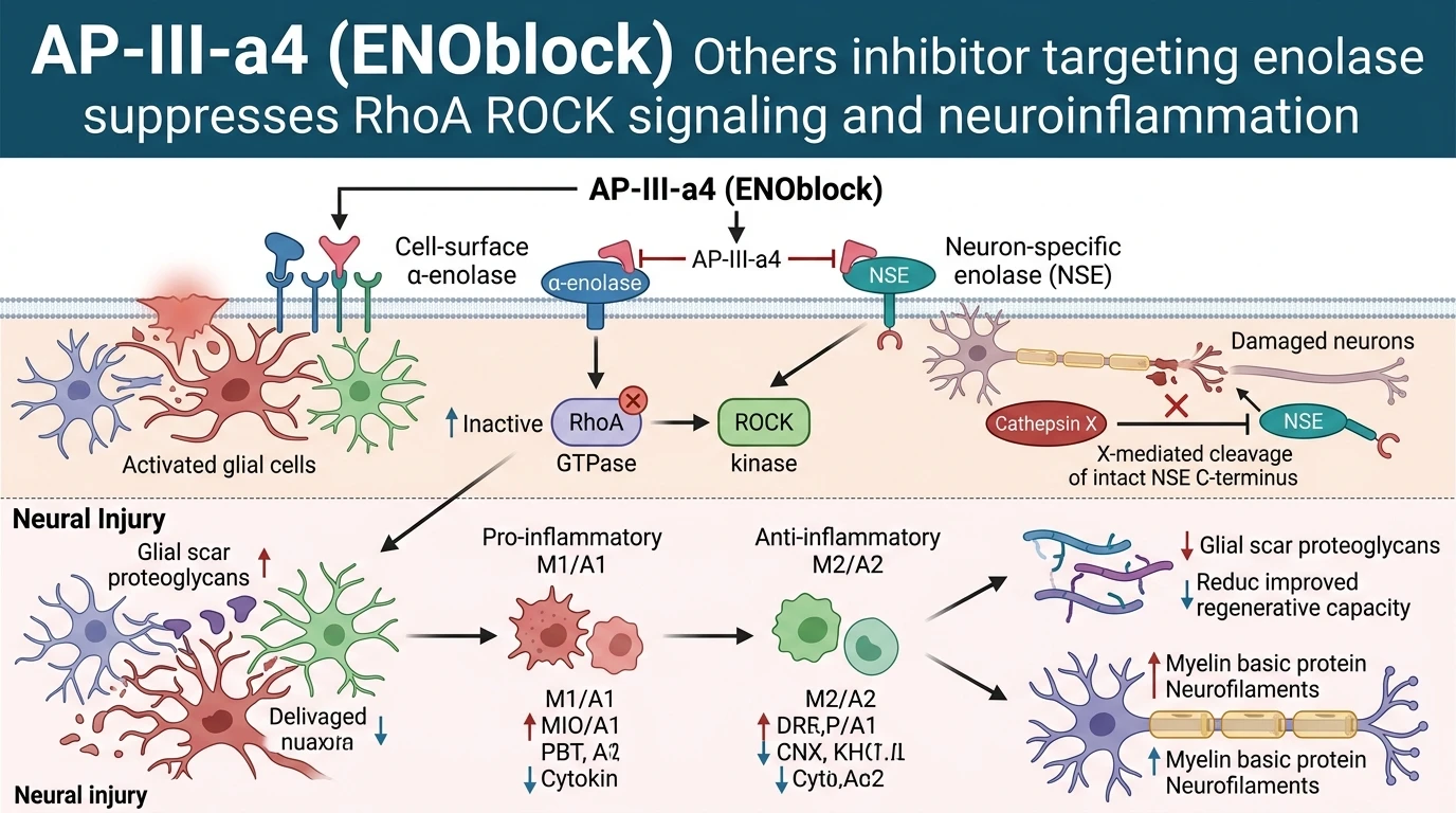
Enolase exists in mammalian systems as homodimeric and heterodimeric isoenzymes formed by α, β, and γ subunits. Under unperturbed metabolic conditions, cytosolic α-enolase (ENO1) and neuron-specific γ-enolase (NSE/ENO2) perform the reversible dehydration of 2-phospho-D-glycerate to phosphoenolpyruvate within the glycolytic pathway. However, physical trauma, ischemia, or neurodegenerative stress induces cellular translocation of these enolase isoforms to the outer leaflet of the cell membrane. On the cell surface, enolase functions as a non-catalytic receptor that regulates extracellular matrix remodeling and inflammatory cell recruitment. Surface enolase accumulation in spinal cord injury and neurodegenerative models correlates with progressive secondary damage, chronic neuroinflammation, and delayed axonal recovery.
The small molecule AP-III-a4 (ENOblock) was discovered as a non-substrate analog inhibitor engineered to disrupt cell-surface enolase interactions. Traditional substrate-competitive enolase inhibitors often induce systemic cytotoxicity by indiscriminately suppressing intracellular glycolytic flux across healthy tissues. In contrast, AP-III-a4 (ENOblock) binds to α-enolase and γ-enolase isoforms, blocking their surface-associated signaling functions. At micromolar working concentrations in vitro and in rodent tissue models, the compound attenuates surface enolase activity without causing metabolic disruption in uninjured cell populations. Target engagement prevents enolase-dependent assembly of pro-inflammatory complexes on activated microglia and reactive astrocytes.
Downstream of surface enolase engagement, intracellular signal transduction relies heavily on the RhoA/Rho-associated protein kinase (ROCK) signaling pathway. Membrane enolase activation stimulates RhoA GTP-binding, initiating ROCK-mediated phosphorylation of downstream effectors including LIM kinase and myosin light chain. This cascade triggers actin filament reorganization, growth cone collapse, and neurite retraction in injured neurons. In primary neuronal cultures and rodent spinal cord injury models, treatment with AP-III-a4 (ENOblock) suppresses RhoA GTP loading and abolishes ROCK pathway activation. Inhibiting this signaling axis prevents cytoskeletal collapse, preserving structural neurofilament integrity and enabling axonal elongation across injury borders.
In parallel with RhoA/ROCK suppression, enolase inhibition impacts Cathepsin X activity. Cathepsin X is a cysteine carboxypeptidase upregulated during acute central nervous system inflammation. Cathepsin X cleaves two C-terminal amino acids from neuron-specific enolase, destroying the intrinsic neurotrophic and survival-promoting properties of intact NSE. Application of AP-III-a4 (ENOblock) decreases both mRNA expression and catalytic carboxypeptidase activity of Cathepsin X in compromised neural tissue. Preserving the intact C-terminus of NSE maintains endogenous neuroprotective signaling cascades. Intact NSE sustains downstream survival pathways, reducing cleavage of executioner caspase-3 and mitigating apoptotic neuronal death.
Targeting surface enolase also alters the neuroinflammatory microenvironment by shifting glial activation states. Microglia and astrocytes exposed to traumatic injury rapidly adopt neurotoxic M1 and A1 phenotypes, releasing pro-inflammatory cytokines such as TNF-α, IL-1β, and IL-6. In rodent models of neural trauma, administration of AP-III-a4 (ENOblock) promotes polarization toward anti-inflammatory M2 microglial and protective A2 astrocyte states. This phenotypic transition diminishes toxic cytokine production and suppresses dense chondroitin sulfate proteoglycan deposition within the glial scar. Reduced scarring creates a permissive environment for axonal sprouting. Pharmacodynamic analyses confirm that systemic delivery of AP-III-a4 (ENOblock) maintains therapeutic concentrations across the blood-spinal cord barrier, sustaining target inhibition up to 24 hours post-dose. Western blot analyses of lesion tissue homogenates confirm elevated expression of myelin basic protein and heavy neurofilament subunits, indicating preserved myelin sheath structure and axonal survival.
Therapeutic Evaluation and Pharmacological Characterization
Copy code to add this image to your page
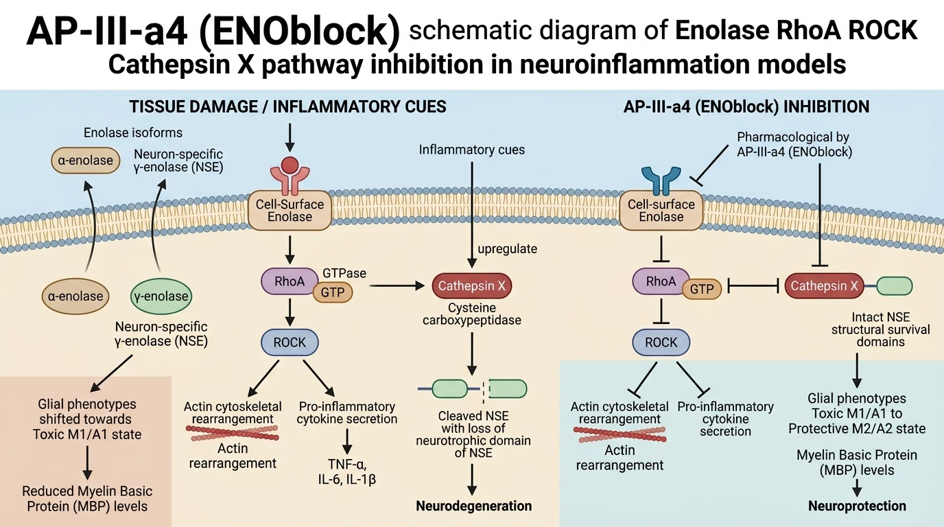
AP-III-a4 (ENOblock) is a small-molecule inhibitor targeting cell-surface α-enolase and neuron-specific γ-enolase (NSE). By inhibiting membrane enolase activation, the compound suppresses downstream RhoA GTPase loading and ROCK kinase activity, preventing axonal cytoskeletal collapse. Concurrently, it suppresses Cathepsin X expression and catalytic activity, preventing C-terminal proteolytic cleavage of NSE to preserve its neurotrophic function. In neural trauma models, AP-III-a4 promotes M2 microglial and A2 astrocyte phenotypes, reduces neurotoxic cytokine expression, attenuates hypertrophic glial scar formation, and elevates myelin basic protein and neurofilament levels, supporting functional neural repair.
RhoA ROCK Cascade Suppression Preserves Cytoskeletal Integrity
In murine models of spinal cord trauma, surface-bound α-enolase acts as a key mediator of neuroinflammatory expansion. Application of AP-III-a4 (ENOblock) Others inhibitor at micromolar concentrations directly interferes with cell-surface enolase activity. This enzymatic blockade limits the GTP-binding activation of RhoA. Downstream ROCK serine/threonine kinase activity drops significantly following exposure. Cytoskeletal collapse in injured axons is markedly reduced. In secondary injury cascades, this mechanism curtails the transcriptional upregulation of pro-inflammatory cytokines including TNF-α and IL-1β. Primary astrocyte cultures treated under hypoxic stress exhibited reduced expression of reactive A1 markers. Concurrent elevation of neuroprotective A2 markers was observed in these cell lines. By halting RhoA GTPase loading, the compound maintains structural axonal stability during acute tissue trauma.
Inhibition of Cathepsin X Cleavage Protects NSE Function
Neuron-specific enolase (NSE) provides critical neurotrophic support through its intact C-terminal peptide domain. Tissue injury induces Cathepsin X upregulation; this carboxypeptidase degrades the C-terminus of NSE and abolishes its pro-survival signalling. Administration of AP-III-a4 (ENOblock) Others inhibitor effectively downregulates both Cathepsin X protein expression and its catalytic activity in rodent neurodegenerative assays. Preserving the structural integrity of the NSE C-terminus retains endogenous neurotrophic cascades. Primary cortical neurons exposed to toxic glutamate concentrations demonstrated enhanced survival metrics when co-incubated with the inhibitor. Neurite outgrowth remained intact despite oxidative challenge. Microglial populations displayed a marked shift from the neurotoxic M1 state toward the anti-inflammatory M2 phenotype. This phenotypic transition coincides with diminished nitric oxide production and enhanced phagocytic clearance of cellular debris.
Attenuation of Glial Scarring and Myelin Maintenance
Spinal cord injury models treated with AP-III-a4 (ENOblock) Others inhibitor demonstrated significant structural preservation within lesion borders. Hypertrophic chondroitin sulfate proteoglycan deposition within the glial scar was quantified as lower in treated animal cohorts compared to vehicle controls. Dense extracellular matrix accumulation typically restricts axonal sprouting. Downregulation of enolase-mediated pathway nodes loosened this physical barrier. Immunoblotting of spinal tissue homogenates revealed elevated levels of myelin basic protein and heavy neurofilament subunits. These biochemical markers confirm attenuated demyelination and structural axonal preservation. Functional motor scores improved steadily in rodent paradigms following systemic compound delivery. These empirical outcomes demonstrate that targeted enolase inhibition mitigates secondary injury mechanisms while creating a permissive environment for neural recovery.