SHP099 Synergizes with Erlotinib to Suppress SHP2-Mediated Signaling in EGFR-Mutant Lung Adenocarcinoma
Copy code to add this image to your page
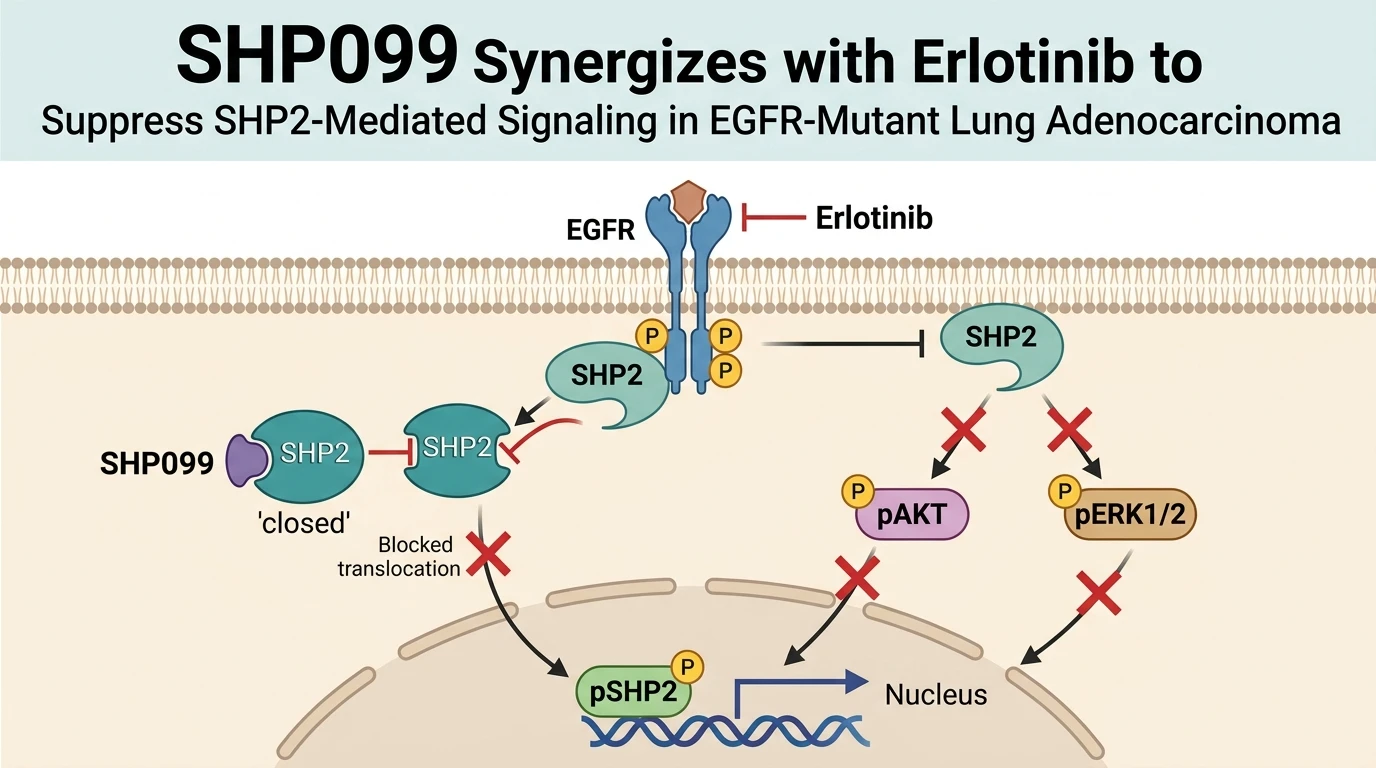
In EGFR mutation-positive lung adenocarcinoma (LUAD), targeted tyrosine kinase inhibitor (TKI) therapies frequently encounter acquired or adaptive resistance. Elevated expression of Src-homology 2 domain-containing phosphatase 2 (SHP2), encoded by the PTPN11 gene, correlates directly with post-resection tumor recurrence. Transcriptomic analyses confirm that high SHP2 mRNA levels predict significantly shorter progression-free survival and overall survival in patients undergoing TKI regimens. At the cellular level, SHP2 acts as a key signal node downstream of activated receptor tyrosine kinases. Exposure to EGFR TKIs such as erlotinib paradoxically increases SHP2 phosphorylation. This phosphorylation induces translocation of pSHP2 from the cytoplasm into the nuclear compartment, where it sustains pro-survival signaling input.
Overcoming this feedback response requires direct intervention at the SHP2 node. The MAPK signaling pathway cascade remains vulnerable to such targeted inhibition. SHP099 acts as an allosteric small-molecule inhibitor that binds at the junction of the N-terminal SH2, C-terminal SH2, and phosphatase domains. This binding event locks SHP2 in an inactive, autoinhibited state. In preclinical models, co-administration of erlotinib with SHP099 effectively prevents the nuclear accumulation of pSHP2. In vitro tests across EGFR-mutant LUAD lines—including PC9, HCC827, and H1975—demonstrate pronounced synergistic suppression of cell viability and clonogenic survival.
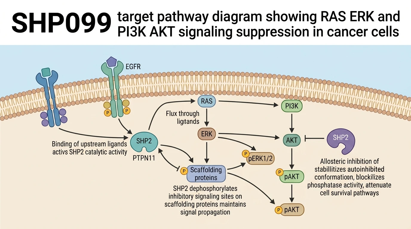
Mechanistically, concurrent blockade of EGFR and SHP2 shuts down critical survival pathways. Erlotinib monotherapy often leaves active residual signal transmission through downstream effectors. Combining erlotinib with SHP099 produces complete suppression of downstream phosphorylation at pAKT (S473) and pERK1/2 (T202/Y204). Short-term cell proliferation and long-term colony formation decline rapidly following dual treatment. Western blot analyses indicate that single-agent erlotinib fails to achieve sustained pERK suppression due to rapid SHP2-mediated feedback loops. Dual target blockade durably inactivates both RAS-ERK and PI3K-AKT signaling cascades, establishing allosteric SHP2 inhibition as a valid strategy to overcome TKI tolerance.
Therapeutic Synergy and Downstream Pharmacological Blockade
Copy code to add this image to your page
SHP099 is a selective, allosteric inhibitor of SHP2 (PTPN11) that locks the phosphatase in its inactive state. In EGFR-mutant lung adenocarcinoma, erlotinib increases SHP2 phosphorylation and drives its nuclear translocation, promoting treatment resistance. Combining erlotinib with SHP099 blocks nuclear accumulation of pSHP2 and completely abrogates downstream pAKT (S473) and pERK1/2 (T202/Y204) activation. This combination yields synergistic suppression of viability and colony formation in EGFR-mutant LUAD cell lines.
Nuclear Translocation Dynamics of Phosphorylated SHP2 Under TKI Challenge
Erlotinib administration rapidly increases tyrosine phosphorylation of cytoplasmic SHP2 in EGFR-mutant lung adenocarcinoma models. Immunofluorescence imaging demonstrates marked nuclear accumulation of pSHP2 following TKI exposure. This subcellular redistribution helps sustain survival signaling despite upstream receptor blockade. Co-treatment with SHP099 prevents this nuclear translocation event. The allosteric inhibitor maintains the closed, inactive conformation of the phosphatase. Consequently, nuclear levels of pSHP2 drop sharply within hours of dual dosing. Cellular assays show that preventing nuclear pSHP2 accumulation correlates directly with heightened sensitivity to erlotinib in PC9 and HCC827 cell lines.
Dual Node Blockade Abrogates Downstream pAKT and pERK Phosphorylation
Single-agent EGFR TKI treatment frequently fails to extinguish downstream effector flux. Combined exposure to erlotinib and SHP099 achieves total clearance of pAKT at residue S473 and pERK1/2 at residues T202/Y204. Immunoblot analyses reveal complete loss of these phospho-epitopes in H1975 cells. Colony formation assays display greater than 80 percent reduction in clonogenic growth under dual therapy compared to single agents. This inhibition occurs at low micromolar concentrations in vitro. Sustained inactivation of both PI3K-AKT and RAS-ERK pathways accounts for the marked synergistic cytotoxicity observed across tested EGFR-mutant models.