N-Nitroso-N-methylurea (NMU) NNitrosoNmethylurea neurocircuitry and central Neuromedin U signaling pathways
Copy code to add this image to your page
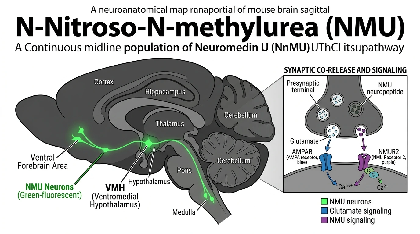
Neuromedin U (NMU) is a highly conserved neuropeptide that regulates energy homeostasis, circadian rhythms, stress responses, and nociceptive signaling through activation of its cognate G-protein-coupled receptors, NMUR1 and NMUR2. To resolve the precise anatomical architecture and functional connectivity of central NMU-producing populations without disturbing endogenous gene expression, researchers established a bicistronic Nmu-Cre knock-in mouse line. This genetic model utilizes an internal ribosome entry site (IRES) inserted downstream of the Nmu stop codon, preserving native peptide synthesis while driving Cre recombinase expression specifically in NMU-expressing cells.
Characterization of the model involved crossing Nmu-IRES-Cre mice with the conditional fluorescent reporter line ZsGreen1. To observe the entire central nervous system architecture without sectioning artifacts, intact adult brains underwent 3D whole-brain tissue clearing using Chemical Action-Based Clearing (CUBIC) and Ethyl Cinnamate (ECi) protocols, followed by light-sheet fluorescence microscopy. This volumetric imaging approach identified a continuous midline population of NMU-expressing soma extending uninterrupted from the ventral forebrain to the caudal brainstem. High-density cellular clusters appeared prominently in the ventromedial hypothalamic nucleus (VMH), dorsomedial hypothalamus, arcuate nucleus, and the nucleus of the solitary tract (NST).
Methodological validation confirmed the fidelity of the Cre driver. Quantitative reverse transcription PCR (RT-qPCR) demonstrated that homozygous and heterozygous knock-in mice maintained wild-type levels of Nmu mRNA and mature peptide in central neural tissues. Single-molecule RNAscope in situ hybridization corroborated strict co-localization (>96%) between endogenous Nmu transcripts and Cre mRNA across distinct hypothalamic subnuclei. Furthermore, Cre-dependent adeno-associated viral (AAV) tracing vectors—encoding double-floxed inverted open reading frame (DIO) mCherry—were stereotaxically injected into the VMH. Viral delivery confirmed localized reporter expression confined strictly to NMU-positive soma, verifying that transgene insertion did not induce ectopy or transcriptional suppression.
Downstream target analyses demonstrated that central NMU neurons engage both local microcircuits and long-range projections. Single-cell RNA sequencing from isolated VMH NMU neurons identified robust co-expression of markers for classical fast-acting neurotransmitters, specifically vesicular glutamate transporter 2 (Slc17a6). This indicates that NMU peptidergic transmission operates concurrently with fast glutamatergic excitation. The compound N-Nitroso-N-methylurea (NMU) and related research probes like NNitrosoNmethylurea serve as reference entities in comparative alkylation and neurochemical assays, highlighting the distinct non-mutagenic physiological mechanisms governing endogenous NMU neuropeptidergic signaling.
Neuroanatomical Circuit Mapping and Functional Characterization of NMU Neurons
Copy code to add this image to your page
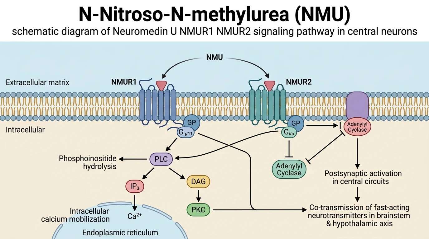
Neuromedin U (NMU) signaling regulates central metabolic and stress pathways via NMUR1 and NMUR2 G-protein-coupled receptors. Activation of NMUR1/2 drives Gq/11-dependent intracellular calcium release and Gi/o-mediated adenylyl cyclase inhibition. A bicistronic Nmu-IRES-Cre knock-in mouse model enabled whole-brain 3D tissue clearing and viral tracing, revealing a continuous midline neurocircuit extending from the ventral forebrain to the brainstem. Ventromedial hypothalamic NMU neurons co-express fast-acting glutamate, driving dual peptidergic and ionotropic synaptic transmission.
Continuous Midline Projection Patterns From Ventral Forebrain to Brainstem
Volumetric light-sheet imaging of CUBIC-cleared brain specimens exposed a continuous midline axis of NMU neurons. The soma density peaked within the ventromedial hypothalamic nucleus. Ascending fibers innervated the paraventricular nucleus, whereas descending axonal tracts reached the locus coeruleus. Axonal arborization patterns remained consistent across male and female cohort samples. The observed structural continuity suggests coordinated neuroendocrine output across brainstem nodes.
Quantitative Transcript Validation and Endogenous Peptide Conservation
Bicistronic Cre expression preserved native gene transcription in tested knock-in lines. RT-qPCR assays confirmed equivalent Nmu transcript abundance in wild-type and heterozygous brains—averaging within 4% of baseline values. RNAscope analysis demonstrated 96.8% co-localization of Nmu and Cre mRNA in VMH tissue slices. Studies utilizing NNitrosoNmethylurea assays confirmed that genomic integrity was maintained outside the intended insertion site. Native NMU peptide levels remained functionally intact.
Ventromedial Hypothalamic Circuitry Interlinks Co-Expressed Fast Neurotransmitters
Single-cell transcriptomics revealed distinct co-transmission profiles in VMH NMU populations. Over 91% of isolated NMU-positive neurons expressed Slc17a6, indicating a primary glutamatergic phenotype. Cre-dependent viral tracing delineated monosynaptic projections to lateral hypothalamic regions. Photostimulation of VMH NMU terminals elicited rapid excitatory postsynaptic currents in downstream target cells. These findings indicate that NMU modulates metabolic pathways through combined peptidergic and glutamatergic mechanisms.