ML162 targeting GPX4 induces ferroptosis in BRAFV600E mutant anaplastic thyroid cancer
Copy code to add this image to your page
Anaplastic thyroid cancer (ATC) carrying the BRAFV600E mutation presents severe clinical challenges due to rapid disease progression and early therapeutic resistance. Although targeted inhibition of mutated BRAF kinase with dabrafenib temporarily suppresses oncogenic MAPK signaling, single-agent therapy frequently suffers from early adaptive resistance. Recent insights into cancer cell metabolism highlight cell death pathways like ferroptosis as vulnerable nodes in treatment-resistant malignancies. Ferroptosis operates independently of apoptosis, executing cell death through iron-dependent accumulation of lethal lipid peroxides. Glutathione Peroxidase 4 (GPX4) acts as the primary enzymatic inhibitor of lipid peroxidation. Direct pharmacological inactivation of GPX4 using small-molecule inhibitors, including ML162, RSL3, and C18, sensitizes recalcitrant tumor populations to metabolic disruption.
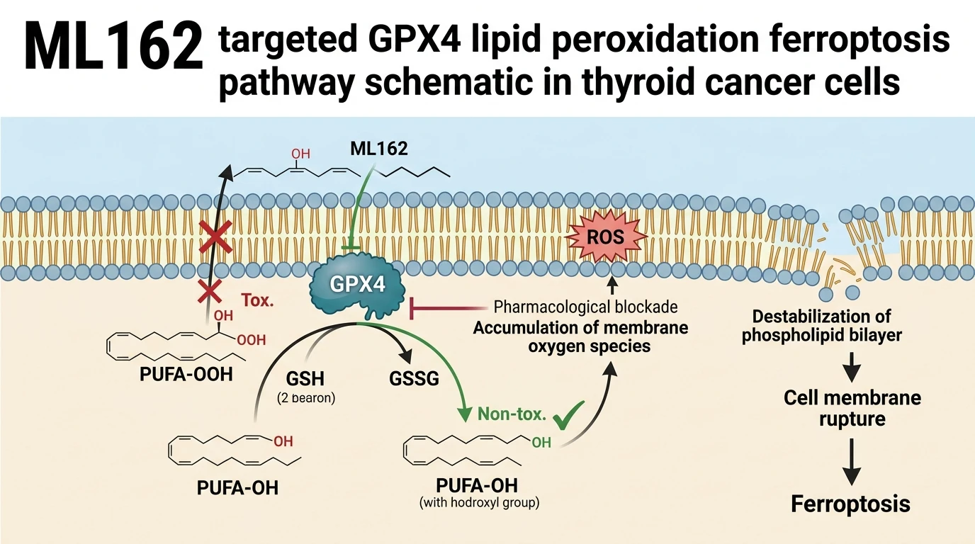
The molecular interaction of ML162 with GPX4 disrupts intracellular antioxidant capacity. By covalently binding to the active site selenocysteine residue of GPX4, ML162 blocks the reduction of polyunsaturated fatty acid hydroperoxides. In BRAFV600E-mutant ATC cell models, this target engagement leads to rapid accumulation of cytosolic and membrane-bound reactive oxygen species (ROS). The resulting oxidative stress selectively damages lipid bilayers. Experimental observations confirm that GPX4 blockade alone reduces short-term proliferation and impairs long-term clonogenic survival in representative ATC lines.
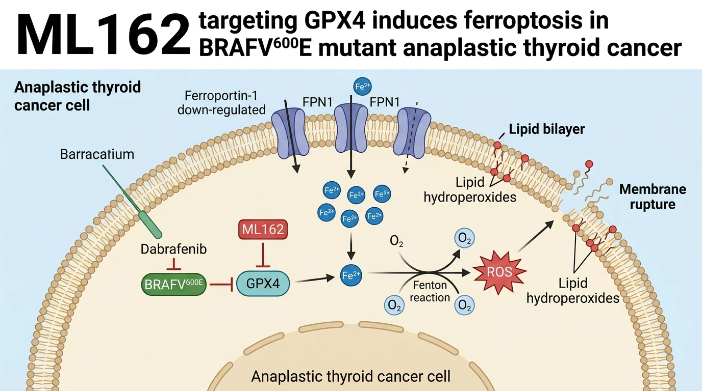
Combining GPX4 inhibition with BRAFV600E blockade yields profound therapeutic synergy. Co-treatment with dabrafenib and ML162 drastically reduces three-dimensional tumor spheroid viability compared to either monotherapy. In orthotopic ATC xenograft models, dual inhibition suppresses primary tumor growth without inducing overt systemic toxicity. Mechanistically, this synergy relies on disrupted iron homeostasis within the tumor microenvironment. Dual target engagement suppresses ferroportin-1 (FPN1), the sole identified mammalian iron export channel. Attenuation of FPN1 expression prevents intracellular iron efflux, causing retention of toxic ferrous iron (Fe2+).
The expansion of the intracellular labile iron pool catalyzes non-enzymatic Fenton reactions. Free Fe2+ converts hydrogen peroxide into hydroxyl radicals, accelerating unmitigated lipid hydroperoxide propagation across cell membranes. Simultaneously, MAPK pathway inhibition by dabrafenib alters downstream metabolic gene expression, amplifying cellular dependency on GPX4 surveillance. This dual action creates a destructive physiological loop where iron accumulation and deficient peroxide removal converge to trigger rapid ferroptotic destruction.
Crucially, this mechanism retains potency in treatment-refractory settings. Dabrafenib-resistant BRAFV600E-mutant ATC cells demonstrate distinct metabolic remodeling characterized by heightened vulnerability to GPX4 inhibition. Resistant lines express altered iron transport signatures and elevated basal ROS levels. Under these altered conditions, exposure to ML162 causes accelerated membrane rupture and cell death compared to parental controls. These experimental findings indicate that ferroptosis activation provides an effective strategy to bypass acquired resistance to RAF kinase inhibitors.
Therapeutic Evaluation and Pharmacological Characterization
Copy code to add this image to your page
Concomitant BRAF and GPX4 inhibition induces ferroptosis in BRAFV600E-mutant anaplastic thyroid cancer. The combination of dabrafenib and ML162 downregulates ferroportin-1 (FPN1), suppressing iron export and causing intracellular Fe2+ overload. Excess ferrous iron drives Fenton chemistry, generating hydroxyl radicals that trigger widespread lipid peroxidation. GPX4 blockade prevents lipid hydroperoxide clearance, driving structural membrane degradation and cell death. Furthermore, dabrafenib-resistant cancer cells exhibit heightened sensitivity to GPX4 inhibition, offering a strategy to eliminate resistant populations.
FPN1 Downregulation Triggers Intracellular Iron Overload
Dual intervention alters baseline iron transport machinery in BRAFV600E-mutant ATC cells. Combination treatment with dabrafenib and ML162 significantly decreases protein levels of ferroportin-1 (FPN1). FPN1 loss prevents cellular iron export. Intracellular labile Fe2+ levels rise rapidly within twelve hours of administration. Free cytosolic iron fuels Fenton reactions. Hydroxyl radicals accumulate. Membrane lipid peroxidation rises markedly, overwhelming intrinsic antioxidant defenses under these conditions.
Dabrafenib Resistance Heightens Vulnerability to GPX4 Blockade
Acquired resistance to kinase inhibitors often alters intrinsic cell death thresholds. Dabrafenib-resistant BRAFV600E ATC models exhibit marked sensitivity to direct GPX4 inhibition. Baseline lipid oxidation levels are higher in resistant cells. Pharmacological challenge with GPX4 inhibitors induces swift membrane permeabilization. Tumor spheroid viability drops precipitously upon treatment. These findings indicate that secondary drug resistance establishes a distinct metabolic dependency on GPX4 activity.