IAG933 Disruption of YAP1-TEAD Complex Suppresses Glioblastoma Vasculogenic Mimicry
Copy code to add this image to your page
Glioblastoma remains among the most refractory primary intracranial malignancies, characterized by pronounced invasive potential, cellular heterogeneity, and persistent therapeutic resistance against standard temozolomide chemotherapies. A major driver of microvascular plasticity and therapeutic evasion in glioblastoma is vasculogenic mimicry (VM), a non-angiogenic fluid microcirculation mechanism wherein aggressive tumor cells transdifferentiate to form matrix-rich, capillary-like tubular structures independently of host endothelial cells. Clinical glioblastoma tissue profiling via immunohistochemistry and RNA sequencing reveals marked upregulation of YAP1 co-activator and its primary transcriptional binding partner TEAD1 relative to non-neoplastic brain tissue control samples. This elevated expression directly correlates with advanced histological tumor grade, increased microvascular density, and reduced overall survival in patient cohorts.
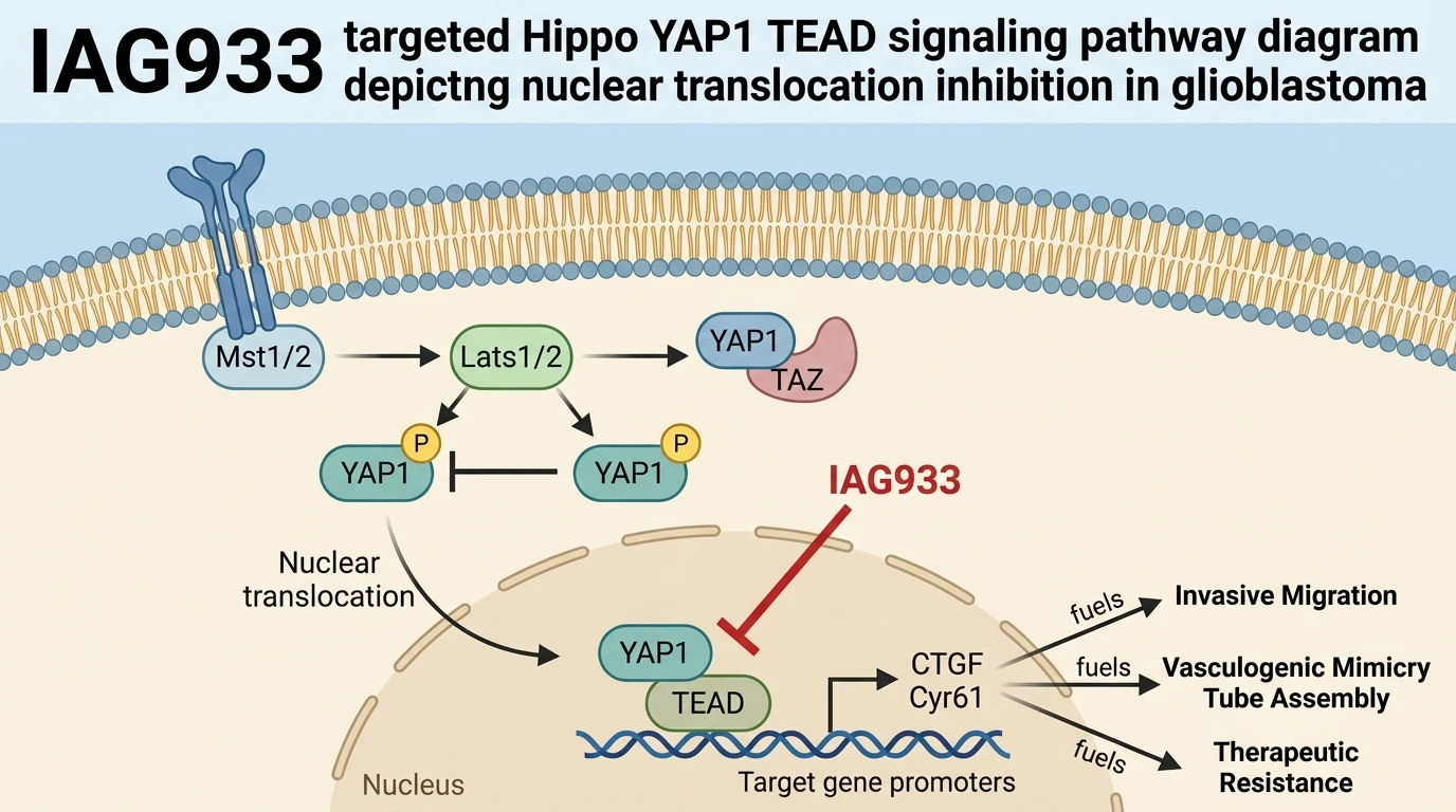
At the biochemical level, the transcription factor complex formed between YAP1 and TEAD domain family members serves as the terminal effector node of the Hippo signaling cascade. Under basal baseline conditions in quiescent glioblastoma cells, active upstream Hippo core kinases—specifically MST1/2 and LATS1/2—phosphorylate YAP1 at canonical residues such as Serine 127. This post-translational modification promotes cytoplasmic retention via 14-3-3 protein binding and targets YAP1 for ubiquitin-mediated proteasomal degradation. However, during functional vasculogenic mimicry induction on basement membrane substrates such as Cultrex extracellular matrix, Hippo cascade flux shifts, permitting YAP1 dephosphorylation and rapid nuclear translocation. Within the nuclear compartment, unphosphorylated YAP1 physically engages TEAD1, forming a functional heterodimeric transcriptional complex. This complex binds promoter elements of target genes, driving robust upregulation of connective tissue growth factor (CTGF) and cysteine-rich angiogenic inducer 61 (Cyr61), two matricellular proteins critical for endothelial-like channel stabilization and cell-matrix interaction.
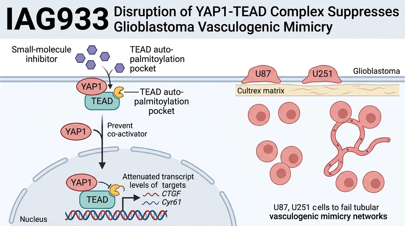
To evaluate the pharmacological vulnerability of this signaling axis, small-molecule disruption of the YAP1-TEAD transcriptional complex was investigated using the direct inhibitor IAG933 alongside comparator Hippo pathway modulators GNE7883 and VT107. In human glioblastoma models—specifically U87 and U251 cell lines—exposure to IAG933 effectively suppressed chemotactic cell migration under Boyden chamber transwell assay conditions. Concurrently, live-cell high-content imaging demonstrated that IAG933 disrupted the assembly of three-dimensional capillary-like VM tube networks on Cultrex matrices in a concentration-dependent manner. Rather than assembling into interconnected, continuous polygonal network loops, IAG933-treated U87 and U251 cells remained as isolated, non-organized spherical cellular clusters.
Mechanistic validation revealed that pharmacological intervention with IAG933 directly altered the subcellular localization kinetics of YAP1. Confocal immunofluorescence microscopy demonstrated that IAG933 exposure attenuated the nuclear accumulation of YAP1 following VM pathway activation, retaining the protein predominantly within the cytosolic compartment. As a direct consequence of compromised nuclear YAP1-TEAD complex formation, quantitative reverse transcription PCR (RT-qPCR) assays confirmed marked suppression of downstream target gene expression, yielding substantial reductions in CTGF and Cyr61 mRNA transcript abundance.
To confirm that the observed cellular phenotypes were mediated specifically by target engagement of the YAP1-TEAD node rather than non-specific compound toxicity, genetic depletion studies were conducted using small interfering RNA (siRNA) directed against YAP1. Genetic silencing of YAP1 closely phenocopied the pharmacodynamic effects observed with IAG933 treatment. Specifically, siRNA-mediated YAP1 depletion led to reduced nuclear YAP1 accumulation, decreased chemotactic migration velocity, complete loss of VM capillary network architecture, and parallel reductions in CTGF and Cyr61 transcript levels.
These empirical observations demonstrate that the Hippo/YAP1-TEAD signaling axis directly coordinates vasculogenic mimicry tube assembly and migratory capacity in glioblastoma cell lines. Targeted disruption using the small-molecule inhibitor IAG933 suppresses these malignant phenotypic properties under tested in vitro assay conditions. By blocking nuclear YAP1-TEAD complex function and downregulating pro-vasculogenic effectors CTGF and Cyr61, small-molecule YAP1-TEAD inhibition represents a validated strategy to suppress microvascular plasticity and target VM-driven chemoresistance in glioblastoma.
Pharmacological Evaluation and Transcriptional Target Engagement
Copy code to add this image to your page
Pharmacological target engagement of the TEAD protein by IAG933 disrupts the assembly of the transcriptional YAP1-TEAD complex. In glioblastoma cell models, this targeted interaction blocks YAP1 nuclear translocation and suppresses downstream transcription of matricellular genes, including CTGF and Cyr61. Consequently, treated cells display impaired chemotactic migration and a reduced capacity to construct three-dimensional capillary-like networks during vasculogenic mimicry on basement membrane matrices. This mechanism offers a targetable biochemical axis to address cell plasticity and chemoresistance.
Inhibition of Chemotactic Migration and Matrix Tube Formation
Assays conducted on U87 and U251 glioblastoma models confirmed that small-molecule treatment with IAG933 impairs functional cell mobility under gradient-driven transwell conditions. Chemotactic migration rates decreased substantially following compound incubation. On Cultrex basement membrane matrices, control cells organized into extensive polygonal capillary-like networks within six hours of seeding. Treatment with the compound disrupted this assembly process. Branch point density dropped significantly across tested concentration ranges; cell clusters failed to establish continuous lumen structures. Similar morphological inhibition was observed with reference inhibitors GNE7883 and VT107. These empirical metrics indicate that small-molecule interference with Hippo signaling directly undermines the structural morphogenesis required for vasculogenic mimicry.
Attenuation of Nuclear YAP1 Translocation and Target Transcription
Subcellular fractionation and immunofluorescence analysis demonstrated that matrix-induced vasculogenic mimicry promotes rapid nuclear import of YAP1 in U87 cells. Direct application of IAG933 reversed this nuclear accumulation, maintaining YAP1 within the cytoplasm. Consequently, transcriptional assembly at TEAD domain promoter sites was restricted. Quantitative PCR assays revealed marked down-regulation of CTGF mRNA levels within twelve hours of drug exposure. Cyr61 transcript abundance exhibited parallel reductions across treated cohorts. siRNA knockdown of YAP1 confirmed this mechanism; genetic suppression yielded equivalent transcriptional loss and tube network collapse. These findings demonstrate that targeted disruption of YAP1-TEAD-dependent transcriptional signaling blocks vasculogenic microvascular adaptation in glioblastoma cell lines.