Ibrutinib (PCI-32765) Inhibits BTK and Downstream Akt/mTOR Signaling in Glioblastoma Models
Copy code to add this image to your page
Bruton's tyrosine kinase (BTK) functions as a non-receptor tyrosine kinase essential for immune cell development and oncogenic signaling cascades. While historically investigated in hematologic malignancies, emerging empirical evidence demonstrates aberrant expression and functional dependency on BTK in solid tumors, including glioblastoma (GBM). In patient-derived GBM primary cells and established cell lines, pharmacological inhibition of BTK enzymatic activity via the small-molecule inhibitor Ibrutinib (PCI-32765) disrupts critical intracellular survival networks. Under tested in vitro assay conditions, selective target engagement halts tumor expansion through distinct cell-autonomous mechanisms.
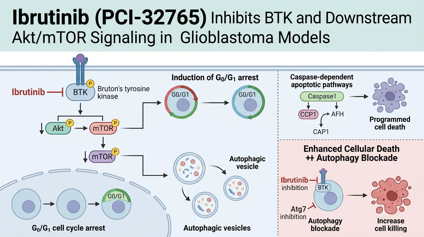
Treatment with Ibrutinib (PCI-32765) induces robust dose- and time-dependent growth inhibition across tested glioblastoma populations. Flow cytometric analysis demonstrates a pronounced G0/G1 phase arrest. This cytostatic response corresponds with down-regulation of cyclin D1 and cyclin-dependent kinase 4 (CDK4), alongside elevated expression of p21 Waf1/Cip1 cyclin-dependent kinase inhibitors. Concurrently, target engagement impairs directional cell migration and invasive capacity in wound-healing and transwell assays. Western blot profiling confirms that catalytic inhibition of BTK triggers annexin V positivity, poly(ADP-ribose) polymerase (PARP) cleavage, and activation of executioner caspase-3, confirming classic apoptotic induction.
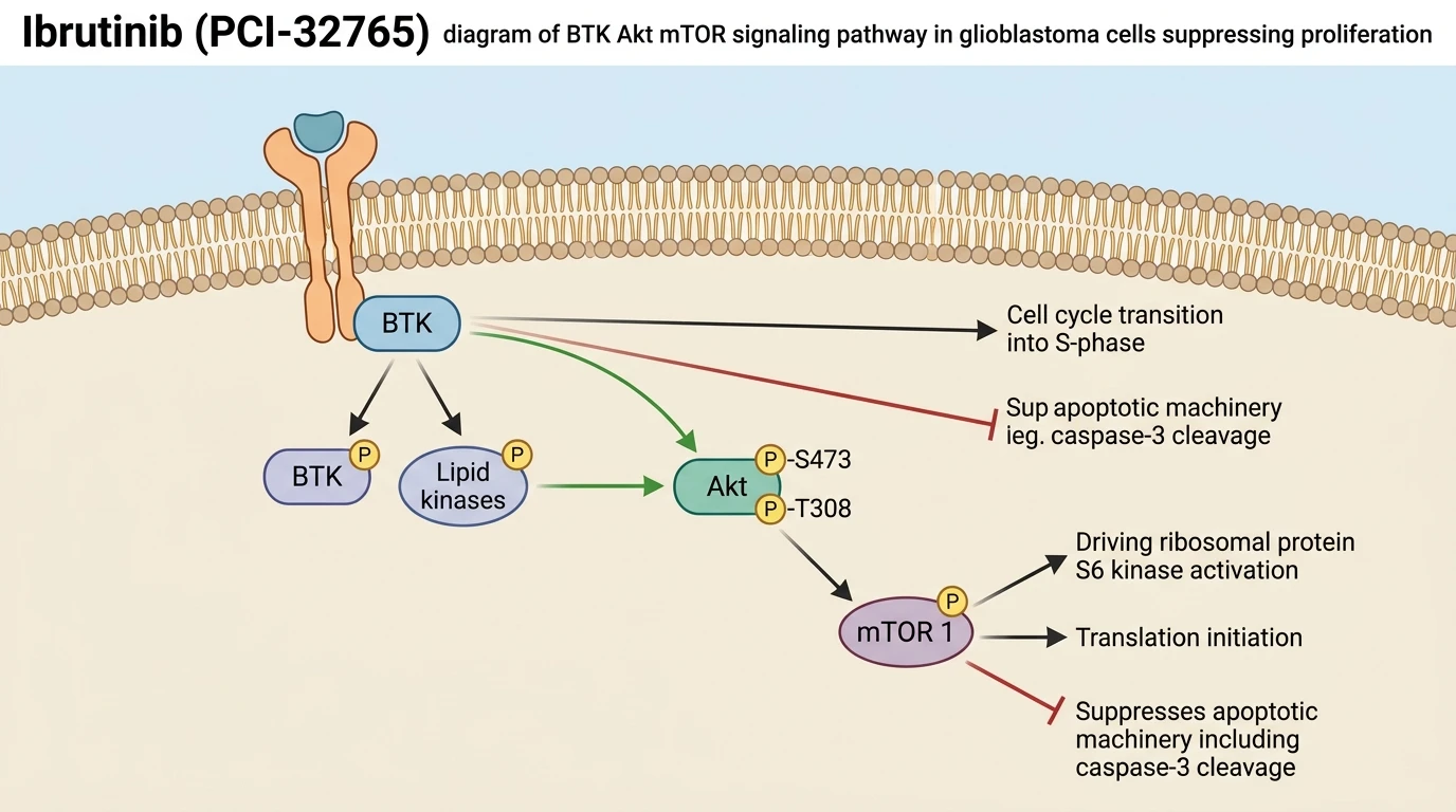
Biochemical pathway mapping reveals that BTK inhibition directly dampens the downstream Akt/mTOR axis. Under basal culture conditions, active BTK maintains phosphorylation of Akt at Ser473. Exposure to Ibrutinib (PCI-32765) abolishes this phosphorylation event within hours. Reduced Akt catalytic activity diminishes phosphorylation of mammalian target of rapamycin (mTOR) and its downstream substrate p70S6 kinase. Suppression of mTOR complex 1 unleashes autophagic flux in glioblastoma cells. This process is documented by increased microtubule-associated protein 1 light chain 3 (LC3-II) conversion and p62 degradation.
Mechanistic assays confirm that the induced autophagy serves a cytoprotective function. Rather than accelerating autophagic cell death, activation of LC3-II sequestration dampens the cytotoxic potential of BTK monotherapy. Genetic disruption of autophagic machinery via Atg7 short hairpin RNA (shRNA) knockdown markedly potentiates drug sensitivity. Similarly, pharmacological blockade using 3-methyladenine (3-MA) enhances apoptotic cleavage markers. Within subcutaneous and orthotopic murine xenograft models, co-administration of autophagy inhibitors with BTK blockade produces superior tumor regression compared to monotherapy. These preclinical observations indicate that dual target engagement represents a potential therapeutic vulnerability in glioblastoma.
Pharmacological Evaluation and Targeted Pathway Blockade
Copy code to add this image to your page
Ibrutinib selectively inhibits Bruton's tyrosine kinase (BTK), disrupting downstream Akt/mTOR pathway signaling in glioblastoma cells. Target engagement inhibits cellular proliferation and cell migration while inducing G0/G1 cell cycle arrest and caspase-mediated apoptosis. Concurrently, suppression of the Akt/mTOR axis induces cytoprotective autophagy. Blocking this autophagic response via chemical inhibitors or Atg7 knockdown enhances apoptotic cell death and antitumor efficacy.
Attenuation of Akt Phosphorylation and G0/G1 Cell Cycle Arrest
At physiologically relevant micromolar concentrations, Ibrutinib (PCI-32765) suppressed downstream phosphorylation of Akt at Ser473 in patient-derived glioblastoma cultures. Downstream phosphorylation ceased within hours. Attenuation of Akt activity diminished mTOR signaling capacity, leading to reduced p70S6K activation—a central node in protein translation and cell proliferation. Cell cycle profiling revealed a substantial accumulation of cells in the G0/G1 phase. Expression levels of cyclin D1 dropped rapidly following exposure. Concurrently, expression of p21 Waf1/Cip1 increased, reinforcing cell cycle arrest. Flow cytometry assays confirmed elevated annexin V staining, indicative of caspase-dependent apoptotic activation in treated populations.
In Vivo Growth Suppression in Murine Xenograft Models
In tumor growth assays within subcutaneous and orthotopic murine xenograft models, daily administration of Ibrutinib (PCI-32765) significantly reduced bioluminescence and tumor volume compared to vehicle controls. Tumor progression slowed dramatically. Immunohistochemical staining of harvested xenograft tissue confirmed reduced Ki-67 proliferation indices and decreased phosphorylated Akt levels. Selectivity margins remained intact without apparent systemic toxicity or significant body weight loss in treated animals. These in vivo observations confirm target engagement and sustained pharmacodynamic suppression of BTK signaling within intracranial lesions under tested physiological conditions.
Autophagy Blockade Enhances Apoptotic Sensitivity in Glioblastoma
Inhibition of BTK-mediated Akt/mTOR signaling simultaneously triggered autophagic vesicle formation within glioblastoma cells. Transmission electron microscopy confirmed double-membrane autophagosome accumulation. To determine whether this response supported survival or cell death, researchers combined BTK inhibition with autophagy blockage. Pharmacological co-treatment with 3-methyladenine or genetic silencing via Atg7 knockdown shifted the cellular outcome from survival to cell death. Cleaved PARP levels increased sharply under dual treatment conditions. Combination strategies effectively converted a cytoprotective feedback response into potent tumor regression within experimental models.