Vadimezan (DMXAA) targeted cGAS-STING signaling regulation and hepatocyte apoptotic responses
Copy code to add this image to your page
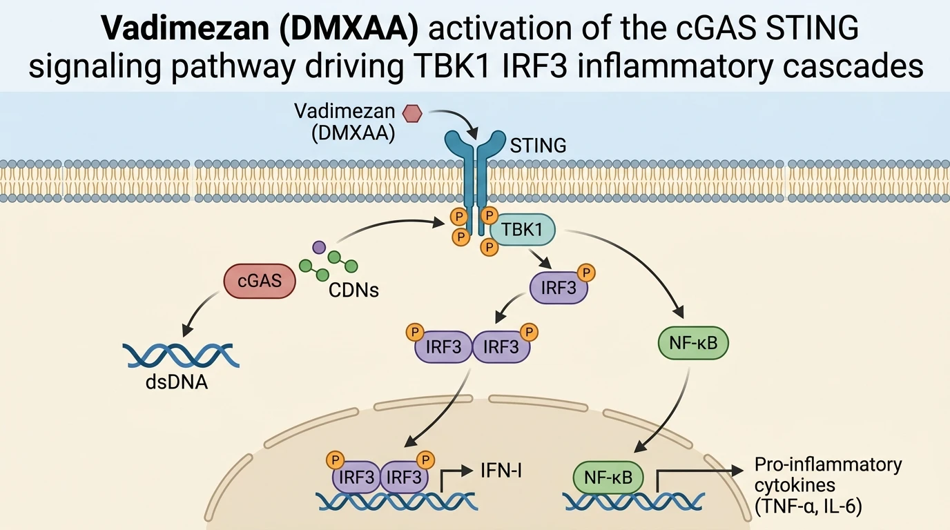
The cyclic GMP-AMP synthase (cGAS)-stimulator of interferon genes (STING) pathway constitutes a central axis in host defense and sterile inflammatory signaling. Activation begins when cGAS detects aberrant cytosolic double-stranded DNA, synthesizing cyclic GMP-AMP (cGAMP) to bind the endoplasmic reticulum-bound receptor STING. Once engaged, STING oligomerizes and translocates to the Golgi apparatus, recruiting TANK-binding kinase 1 (TBK1). This recruitment enables phosphorylation of both STING and interferon regulatory factor 3 (IRF3), alongside activation of nuclear factor kappa B (NF-κB). Downstream signaling triggers robust transcription of type I interferons (IFN-α/β) and pro-inflammatory cytokines, including TNF-α and IL-6. While essential for anti-pathogen immunity, chronic or hyperactive cGAS-STING activation precipitates severe tissue injury, inflammatory cascades, and cell loss across toxicological settings such as acetaminophen-induced acute liver injury (AILI).
In murine systems, Vadimezan (DMXAA) serves as a selective small-molecule STING agonist. Direct binding of Vadimezan induces conformational remodeling of mouse STING, initiating robust downstream phosphorylation of TBK1 and IRF3. Consequently, treatment with Vadimezan elevates local production of IFN-I and inflammatory cytokines in primary macrophages and tissue models. This pharmacological property makes Vadimezan a key tool compound for probing STING-dependent pathways in rodents, despite its species-specific binding selectivity that limits direct engagement with human STING variants.
In primary hepatocytes and Kupffer cells, APAP challenge induces rapid release of mitochondrial DNA (mtDNA) into the cytosol. Cytosolic mtDNA engages cGAS, elevating intracellular cGAMP levels. Subcellular localization studies demonstrate that activated STING undergoes trafficking from the endoplasmic reticulum to perinuclear Golgi compartments. There, STING serves as a scaffolding protein for TBK1 dimerization. Phosphorylated TBK1 then transphosphorylates the C-terminal tail of STING, creating a docking platform for IRF3 recruitment. Subsequent IRF3 phosphorylation leads to homodimerization and nuclear translocation.
When Vadimezan is applied to murine cell culture assays, it bypasses upstream cGAS activation by directly binding the ligand-binding domain (LBD) of murine STING. The compound induces a closed dimeric conformation that recruits TBK1 with high affinity. This triggers immediate phosphorylation of IRF3 within 30 to 60 minutes of administration. In murine models, administration of Vadimezan mimics the sterile inflammatory pathology observed during severe hepatic damage, inducing hepatic neutrophil infiltration and oxidative stress markers.
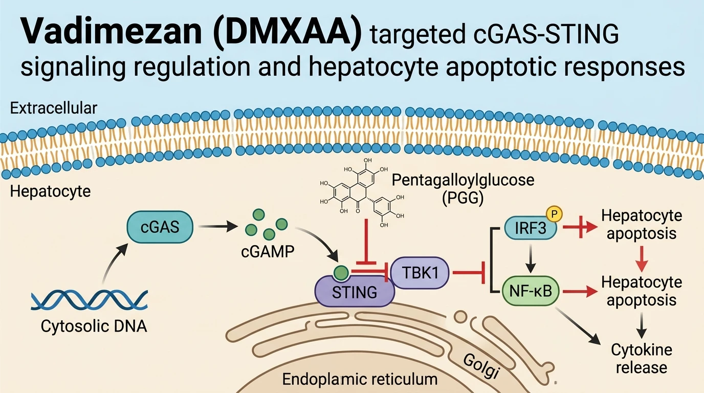
Pentagalloylglucose (PGG), a naturally occurring polyphenolic compound, functions as a broad-spectrum inhibitor of the cGAS-STING cascade. Detailed molecular investigations demonstrate that PGG suppresses both acetaminophen- and Vadimezan-induced inflammatory responses across in vitro and in vivo models. Mechanistically, PGG blocks the physical binding interaction between STING and TBK1, preventing STING-TBK1 complex assembly. Interruption of this protein-protein interface halts downstream IRF3 and NF-κB activation, effectively blocking hepatocyte apoptosis, lipid peroxidation, and oxidative damage. Functional characterization of enzyme kinetics and target interactions can be referenced in established enzymatic data repositories, confirming the distinct binding behavior of polyphenolic modulators within kinase signaling complexes.
In vivo evaluation using APAP-induced murine liver injury models validates these cellular findings. PGG administration markedly reduces serum alanine aminotransferase (ALT) and aspartate aminotransferase (AST) levels relative to vehicle controls. Histopathological examinations reveal preserved hepatic lobular architecture and reduced necrotic foci in PGG-treated mice. Furthermore, PGG attenuates expression of terminal apoptotic markers, including cleaved caspase-3 and TUNEL-positive hepatocytes. These outcomes establish that targeted disruption of the STING-TBK1 signaling node counteracts both drug-induced and agonist-induced hepatic inflammation.
Pharmacological Disruption of the cGAS-STING Signaling Cascade
Copy code to add this image to your page
Vadimezan (DMXAA) acts as a selective murine STING agonist that triggers STING dimerization, TBK1 recruitment, IRF3/NF-κB phosphorylation, and pro-inflammatory cytokine release. In acetaminophen-induced acute liver injury, released mitochondrial DNA similarly hyperactivates cGAS-STING signaling. Pentagalloylglucose (PGG) acts as a broad-spectrum cGAS-STING pathway inhibitor by directly blocking the STING-TBK1 protein-protein interaction. This binding disruption prevents downstream phosphorylation cascades, nuclear translocation of IRF3, hepatocyte apoptosis, and oxidative tissue damage in murine models.
Disruption of STING-TBK1 Association Prevents Downstream IRF3 Phosphorylation
The interaction between STING and TBK1 represents an essential step in cyclic dinucleotide-mediated signaling cascades. At physiologically relevant concentrations in primary murine hepatocytes, PGG directly interferes with STING-TBK1 complex assembly. Western blot analysis confirmed that PGG treatment eliminated STING Ser365 phosphorylation following activation. Downstream IRF3 phosphorylation at Ser396 dropped within thirty minutes. Short-term kinetic measurements demonstrated a parallel decrease in nuclear IRF3 accumulation. Consequently, transcript levels of interferon-beta and interleukin-6 decreased significantly. In murine macrophage assays stimulated with Vadimezan, PGG suppressed pro-inflammatory cytokine expression in a dose-dependent manner. This selective disruption leaves upstream cGAMP synthesis intact while completely arresting signal propagation at the kinase recruitment stage.
Suppression of Hepatocyte Apoptosis and Oxidative Stress in Murine AILI
In vivo validation within an APAP-induced acute liver injury model demonstrates significant hepatoprotection following STING pathway inhibition. Murine subjects receiving high-dose APAP exhibited widespread centrilobular necrosis and elevated serum transaminases. Pretreatment with PGG reduced serum ALT and AST levels by over sixty percent relative to untreated controls. The histological damage score dropped markedly. Mechanistically, PGG administration blocked hepatic malondialdehyde generation and preserved reduced glutathione pools. Cleaved caspase-3 levels remained low in treated liver tissues. When challenging mice with Vadimezan, PGG attenuated systemic release of TNF-alpha and prevented secondary necrotic liver injury. These empirical metrics confirm that disrupting the STING-TBK1 interface limits both acute oxidative damage and inflammatory hepatocyte death.